Parvalbumin Rabbit Polyclonal Antibody

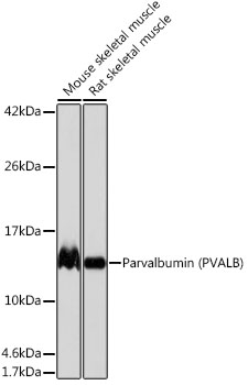
Western blot analysis of various lysates using Parvalbumin (PVALB) Rabbit PAb  at 1:1000 dilution.
Secondary antibody: HRP Goat Anti-Rabbit IgG (H+L) at 1:10000 dilution.
Lysates/proteins: 25μg per lane.
Blocking buffer: 3% nonfat dry milk in TBST.
Detection: ECL West Pico Plus. 
Exposure time: 1s.

Western blot analysis of various lysates using Parvalbumin (PVALB) Rabbit PAb at 1:1000 dilution. Secondary antibody: HRP Goat Anti-Rabbit IgG (H+L) at 1:10000 dilution. Lysates/proteins: 25μg per lane. Blocking buffer: 3% nonfat dry milk in TBST. Detection: ECL West Pico Plus. Exposure time: 1s.

Catalog Number:AB-80654
Conjugate:Unconjugated
Size:100 ug
Concentration:1mg/ml
Host:Rabbit
Isotype:IgG
Clone:POLY
Immunogen:Recombinant fusion protein containing a sequence corresponding to amino acids 1-180 of human Parvalbumin (PVALB).
Reactivity:Human,Mouse,Rat
Applications:Western Blot: 1:500 - 1:1000 Immunohistochemistry(paraffin-embedded tissues): 1:50 - 1:200 Immunofluorescence: 1:50 - 1:200 Immunocytochemistry: 1:50 - 1:200
Molecular Weight:12kDa
Purification:Affinity purification
Form:Liquid
Synonyms:D22S749; Parvalbumin (PVALB)

Applications

Western Blot: 1:500 - 1:1000 Immunohistochemistry(paraffin-embedded tissues): 1:50 - 1:200 Immunofluorescence: 1:50 - 1:200 Immunocytochemistry: 1:50 - 1:200

Immunogen

Recombinant fusion protein containing a sequence corresponding to amino acids 1-180 of human Parvalbumin (PVALB).

Target Background

The protein encoded by this gene is a high affinity calcium ion-binding protein that is structurally and functionally similar to calmodulin and troponin C. The encoded protein is thought to be involved in muscle relaxation. Alternative splicing results in multiple transcript variants.

Synonyms:D22S749; Parvalbumin (PVALB)

Storage

Store at -20℃. Avoid freeze / thaw cycles.

Buffer

PBS with 0.02% sodium azide,0.05% BSA,50% glycerol,pH7.3.

Western blot analysis of various lysates using Parvalbumin (PVALB) Rabbit PAb at 1:1000 dilution. Secondary antibody: HRP Goat Anti-Rabbit IgG (H+L) at 1:10000 dilution. Lysates/proteins: 25μg per lane. Blocking buffer: 3% nonfat dry milk in TBST. Detection: ECL West Pico Plus. Exposure time: 1s.

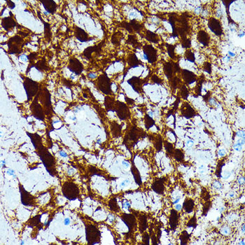
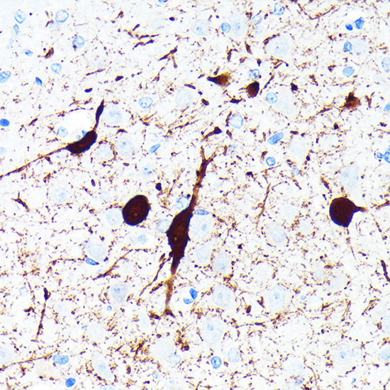
Immunohistochemistry analysis of Parvalbumin (PVALB) in paraffin-embedded mouse brain using Parvalbumin (PVALB) (PVALB) Rabbit pAb at dilution of 1:100 (40x lens).Perform microwave antigen retrieval with 10 mM PBS buffer pH 7.2 before commencing with IHC staining protocol.

Immunohistochemistry analysis of Parvalbumin (PVALB) in paraffin-embedded rat brain using Parvalbumin (PVALB) (PVALB) Rabbit pAb at dilution of 1:100 (40x lens).Perform microwave antigen retrieval with 10 mM PBS buffer pH 7.2 before commencing with IHC staining protocol.

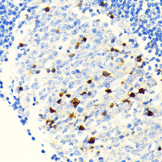
Immunohistochemistry analysis of Parvalbumin (PVALB) in paraffin-embedded human appendix using Parvalbumin (PVALB) (PVALB) Rabbit pAb at dilution of 1:100 (40x lens).Perform microwave antigen retrieval with 10 mM PBS buffer pH 7.2 before commencing with IHC staining protocol.

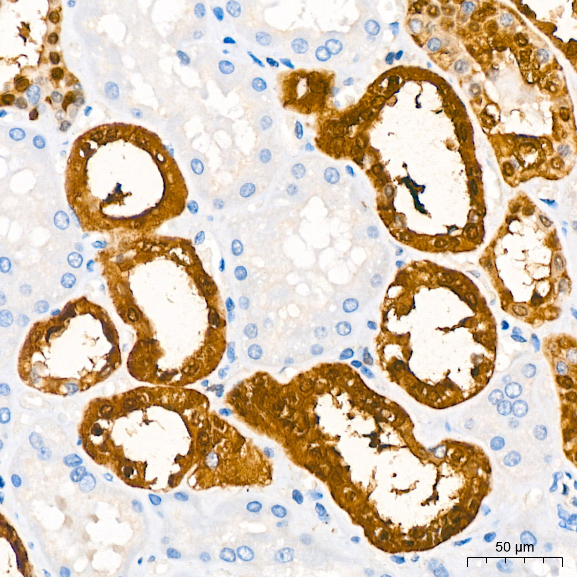
Immunohistochemistry analysis of Parvalbumin (PVALB) in paraffin-embedded human kidney using Parvalbumin (PVALB) Rabbit pAb at dilution of 1:200 (40x lens).Perform high pressure antigen retrieval with 10 mM citrate buffer pH 6.0 before commencing with IHC staining protocol.

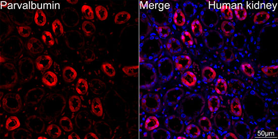
Confocal imaging of paraffin-embedded Human kidney tissue using Parvalbumin (PVALB) Rabbit pAb, dilution 1:100) followed by a further incubation with Cy3 Goat Anti-Rabbit IgG (H+L) dilution 1:500) (Red). DAPI was used for nuclear staining (Blue). High pressure antigen retrieval performed with 0.01M Citrate Buffer(pH 6.0) prior to IF staining. Objective: 40x.

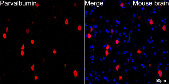
Confocal imaging of paraffin-embedded Mouse brain tissue using Parvalbumin (PVALB) Rabbit pAb (dilution 1:100) followed by a further incubation with Cy3 Goat Anti-Rabbit IgG (H+L) (dilution 1:500) (Red). DAPI was used for nuclear staining (Blue). Microwave antigen retrieval performed with 0.01M Citrate Buffer(pH 6.0) prior to IF staining. Objective: 40x.

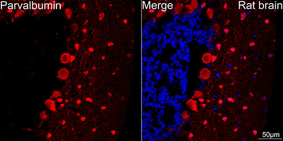
Confocal imaging of paraffin-embedded Rat brain tissue using Parvalbumin (PVALB) Rabbit pAb, dilution 1:100 followed by a further incubation with Cy3 Goat Anti-Rabbit IgG (H+L) (dilution 1:500) (Red). DAPI was used for nuclear staining (Blue). Microwave antigen retrieval performed with 0.01M Citrate Buffer(pH 6.0) prior to IF staining. Objective: 40x.

No documents available.