PD-1 Rabbit Polyclonal Antibody

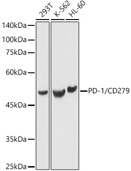
Western blot analysis of various lysates using PD-1/CD279 Rabbit pAb  at 1:1000 dilution.
Secondary antibody: HRP-conjugated Goat anti-Rabbit IgG (H+L)  at 1:10000 dilution.
Lysates/proteins: 25μg per lane.
Blocking buffer: 3% nonfat dry milk in TBST.
Detection: ECL West Pico Plus.
Exposure time: 1s.

Western blot analysis of various lysates using PD-1/CD279 Rabbit pAb  at 1:1000 dilution. Secondary antibody: HRP-conjugated Goat anti-Rabbit IgG (H+L)  at 1:10000 dilution. Lysates/proteins: 25μg per lane. Blocking buffer: 3% nonfat dry milk in TBST. Detection: ECL West Pico Plus. Exposure time: 1s.

Catalog Number:AB-84286
Conjugate:Unconjugated
Size:100 ug
Concentration:1mg/ml
Host:Rabbit
Isotype:IgG
Clone:POLY
Immunogen:Recombinant fusion protein containing a sequence corresponding to amino acids 205-259 of human PD-1/CD279.
Reactivity:Human,Mouse,Rat
Applications:Western Blot:   1:500 - 1:1000 Immunofluorescence: 1:50 - 1:200 Immunocytochemistry: 1:50 - 1:200 ELISA Recommended startingconcentration is 1 μg/mL. Please optimize the concentration based on your specific assay requirements.
Molecular Weight:50kDa/35kDa
Purification:Affinity purification
Form:Liquid
Synonyms:PD1; PD-1; CD279; SLEB2; hPD-1; hPD-l; hSLE1; PD-1/CD279

Applications

Western Blot:   1:500 - 1:1000 Immunofluorescence: 1:50 - 1:200 Immunocytochemistry: 1:50 - 1:200 ELISA Recommended startingconcentration is 1 μg/mL. Please optimize the concentration based on your specific assay requirements.

Immunogen

Recombinant fusion protein containing a sequence corresponding to amino acids 205-259 of human PD-1/CD279.

Target Background

Programmed cell death protein 1 (PDCD1) is an immune-inhibitory receptor expressed in activated T cells; it is involved in the regulation of T-cell functions, including those of effector CD8+ T cells. In addition, this protein can also promote the differentiation of CD4+ T cells into T regulatory cells. PDCD1 is expressed in many types of tumors including melanomas, and has demonstrated to play a role in anti-tumor immunity. Moreover, this protein has been shown to be involved in safeguarding against autoimmunity, however, it can also contribute to the inhibition of effective anti-tumor and anti-microbial immunity.

Synonyms:PD1; PD-1; CD279; SLEB2; hPD-1; hPD-l; hSLE1; PD-1/CD279

Storage

Store at -20℃. Avoid freeze / thaw cycles.

Buffer

PBS with 0.02% sodium azide,50% glycerol,pH7.3.

Western blot analysis of various lysates using PD-1/CD279 Rabbit pAb  at 1:1000 dilution. Secondary antibody: HRP-conjugated Goat anti-Rabbit IgG (H+L)  at 1:10000 dilution. Lysates/proteins: 25μg per lane. Blocking buffer: 3% nonfat dry milk in TBST. Detection: ECL West Pico Plus. Exposure time: 1s.

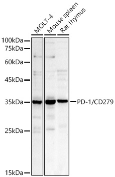
Western blot analysis of various lysates using PD-1/CD279 Rabbit pAb  at 1:1000 dilution. Secondary antibody: HRP-conjugated Goat anti-Rabbit IgG (H+L) at 1:10000 dilution. Lysates / proteins: 25 μg per lane. Blocking buffer: 3 % nonfat dry milk in TBST. Detection: ECL West Pico Plus. Exposure time: 30s.

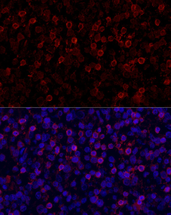
Immunofluorescence analysis of paraffin-embedded human tonsil using PD-1/CD279 Rabbit pAb  at dilution of 1:100 (40xlens). Secondary antibody: Cy3- conjugated Goat anti-Rabbit IgG(H+L) (AS007) at 1:500 dilution. Blue: DAPI for nuclear staining.

No documents available.