PD1 Mouse Monoclonal Antibody

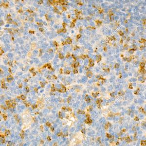
IHC analysis of PD1.Sample: Human tonsil, 4% PFA 12-24h.Antigen retrieval: Citrate buffer , pressure cooker 2 min.Primary antibody: 1: 1200, 4℃ overnight.Secondary antibody: HRP conjugated Goat Anti-Mouse lgG , 1: 200, RT, 1h.

IHC analysis of PD1.Sample: Human tonsil, 4% PFA 12-24h.Antigen retrieval: Citrate buffer , pressure cooker 2 min.Primary antibody: 1: 1200, 4℃ overnight.Secondary antibody: HRP conjugated Goat Anti-Mouse lgG , 1: 200, RT, 1h.

Catalog Number:MABS12338
Conjugate:unconjugated
Size:100 μL
Concentration:1mg/ml
Host:Rabbit
Isotype:IgG1, κ
Clone:IS-11
Immunogen:KLH conjugated Synthetic peptide corresponding to Mouse PD1
Reactivity:Species, Human
Applications:Applications Dilution<br/>IHC/IF 1: 400-1: 2400
Purification:Affinity purification
Form:Liquid
Synonyms:Protein PD-1, mPD-1, CD279, Pdcd1, Pd1, SLEB2, Systemic lupus erythematosus susceptibility 2, hSLE1, PDCD1,PD-1, PD1, SLEB2, hPD-1, hPD-l, Programmed cell death 1

Programmed cell death 1 (PD-1, also known as CD279) is an immunoinhibitory receptor that belongs to the CD28/CTLA-4 subfamily of the Ig superfamily. It is a 288 amino acid (aa) type I transmembrane protein composed of one Ig superfamily domain, a stalk, a transmembrane domain, and an intracellular domain containing an immunoreceptor tyrosine-based inhibitory motif (ITIM) as well as an immunoreceptor tyrosine-based switch motif (ITSM) . PD-1 is expressed during thymic development and is induced in a variety of hematopoietic cells in the periphery by antigen receptor signaling and cytokines.

Applications

Applications Dilution<br/>IHC/IF 1: 400-1: 2400

Immunogen

KLH conjugated Synthetic peptide corresponding to Mouse PD1
Target Background

Programmed cell death 1 (PD-1, also known as CD279) is an immunoinhibitory receptor that belongs to the CD28/CTLA-4 subfamily of the Ig superfamily. It is a 288 amino acid (aa) type I transmembrane protein composed of one Ig superfamily domain, a stalk, a transmembrane domain, and an intracellular domain containing an immunoreceptor tyrosine-based inhibitory motif (ITIM) as well as an immunoreceptor tyrosine-based switch motif (ITSM) . PD-1 is expressed during thymic development and is induced in a variety of hematopoietic cells in the periphery by antigen receptor signaling and cytokines.

Synonyms:Protein PD-1, mPD-1, CD279, Pdcd1, Pd1, SLEB2, Systemic lupus erythematosus susceptibility 2, hSLE1, PDCD1,PD-1, PD1, SLEB2, hPD-1, hPD-l, Programmed cell death 1

Storage

Store at -20℃ for one year. Avoid repeated freeze/thaw cycles.

Buffer

PBS with 0.15% ProClin300, 100 μg/mL BSA and 50% glycerol.

IHC analysis of PD1.Sample: Human tonsil, 4% PFA 12-24h.Antigen retrieval: Citrate buffer , pressure cooker 2 min.Primary antibody: 1: 1200, 4℃ overnight.Secondary antibody: HRP conjugated Goat Anti-Mouse lgG , 1: 200, RT, 1h.

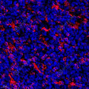
IF analysis of PD1red.Sample: Human B-cell lymphoma, 4% PFA 12-24h.Antigen retrieval: Citrate buffer , pressure cooker 2 min.Blocking buffer: 3% BSA in PBS, RT, 30min.Primary antibody: 1: 400, 4℃ overnight.Secondary antibody: Cy3 conjugated Goat Anti-Mouse IgG , 1: 300, RT, 1h.

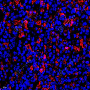
IF analysis of PD1red.Sample: Human tonsil, 4% PFA 12-24h.Antigen retrieval: Citrate buffer , pressure cooker 2 min.Blocking buffer: 3% BSA in PBS, RT, 30min.Primary antibody: 1: 400, 4℃ overnight.Secondary antibody: Cy3 conjugated Goat Anti-Mouse IgG , 1: 300, RT, 1h.

No documents available.