PDGFR Alpha Rabbit Polyclonal Antibody

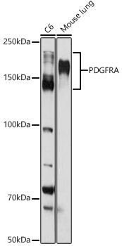
Western blot analysis of extracts of various cell lines, using PDGFR alpha antibody at 1:1000
dilution.
Secondary antibody: HRP Goat Anti-Rabbit IgG (H+L) at 1:10000 dilution.
Lysates/proteins: 25μg per lane.
Blocking buffer: 3% nonfat dry milk in TBST.
Detection: ECL West Pico Plus.
Exposure time: 30s.

Western blot analysis of extracts of various cell lines, using PDGFR alpha antibody at 1:1000 dilution. Secondary antibody: HRP Goat Anti-Rabbit IgG (H+L) at 1:10000 dilution. Lysates/proteins: 25μg per lane. Blocking buffer: 3% nonfat dry milk in TBST. Detection: ECL West Pico Plus. Exposure time: 30s.

Catalog Number:AB-84772
Conjugate:Unconjugated
Size:100 ug
Concentration:1mg/ml
Host:Rabbit
Isotype:IgG
Clone:POLY
Immunogen:Recombinant fusion protein containing a sequence corresponding to amino acids 896-1059 of human PDGFR alpha
Reactivity:Human, Mouse, Rat
Applications:Western Blot: 1:500 - 1:1000 Immunohistochemistry( paraffin embedded tissues) 1:50 - 1:200 Immunofluorescence: 1:50 - 1:200 Immunocytchemistry: 1:50 - 1:200
Molecular Weight:150-190kDa
Purification:Affinity purification
Form:Liquid
Synonyms:CD140A; PDGFR2; PDGFR-2

Applications

Western Blot: 1:500 - 1:1000 Immunohistochemistry( paraffin embedded tissues) 1:50 - 1:200 Immunofluorescence: 1:50 - 1:200 Immunocytchemistry: 1:50 - 1:200

Immunogen

Recombinant fusion protein containing a sequence corresponding to amino acids 896-1059 of human PDGFR alpha

Target Background

This gene encodes a cell surface tyrosine kinase receptor for members of the plateletderived growth factor family. These growth factors are mitogens for cells of mesenchymal origin. The identity of the growth factor bound to a receptor monomer determines whether the functional receptor is a homodimer or a heterodimer, composed of both platelet-derived growth factor receptor alpha and beta polypeptides. Studies suggest that this gene plays a role in organ development, wound healing, and tumor progression. Mutations in this gene have been associated with idiopathic hypereosinophilic syndrome, somatic and familial gastrointestinal stromal tumors, and a variety of other cancers.

Synonyms:CD140A; PDGFR2; PDGFR-2

Storage

Store at -20℃. Avoid freeze / thaw cycles.

Buffer

PBS with 0.02% sodium azide,50% glycerol,pH7.3.

Western blot analysis of extracts of various cell lines, using PDGFR alpha antibody at 1:1000 dilution. Secondary antibody: HRP Goat Anti-Rabbit IgG (H+L) at 1:10000 dilution. Lysates/proteins: 25μg per lane. Blocking buffer: 3% nonfat dry milk in TBST. Detection: ECL West Pico Plus. Exposure time: 30s.

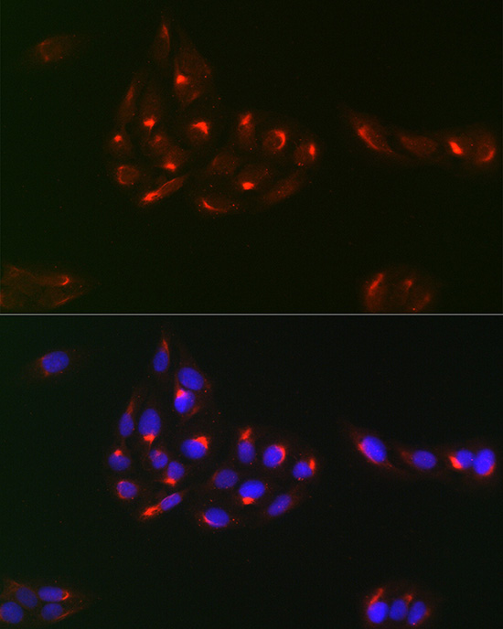
Immunofluorescence analysis of U2OS using PDGFR antibody at dilution of 1:100 (40x lens). Blue: DAPI for nuclear staining.

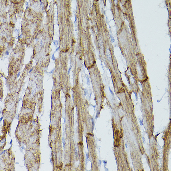
Immunohistochemistry analysis of paraffin-embedded mouse heart using PDGFR alpha Rabbit pAb at dilution of 1:50 (40x lens).Perform high pressure antigen retrieval with 10 mM citrate buffer pH 6.0 before commencing with IHC staining protocol.

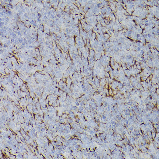
Immunohistochemistry analysis of paraffin-embedded rat spleen using PDGFR alpha Rabbit pAb at dilution of 1:50 (40x lens).Perform high pressure antigen retrieval with 10 mM citrate buffer pH 6.0 before commencing with IHC staining protocol.

No documents available.