PDGFRα(7A3)Mouse Monoclonal Antibody

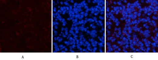
Immunofluorescence analysis of mouse-spleen tissue. 1,PDGFRα Mouse Monoclonal Antibody(7A3)(red) was diluted at 1:200(4°C,overnight). 2, Cy3 labled Secondary antibody was diluted at 1:300(room temperature, 50min).3, Picture B: DAPI(blue) 10min. Picture A:Target. Picture B: DAPI. Picture C: merge of A+B

Immunofluorescence analysis of mouse-spleen tissue. 1,PDGFRα Mouse Monoclonal Antibody(7A3)(red) was diluted at 1:200(4°C,overnight). 2, Cy3 labled Secondary antibody was diluted at 1:300(room temperature, 50min).3, Picture B: DAPI(blue) 10min. Picture A:Target. Picture B: DAPI. Picture C: merge of A+B

Catalog Number:MABN15907
Conjugate:Unconjugated
Size:100ul
Concentration:1mg/ml
Host:Mouse
Isotype:IgG
Clone:7A3
Immunogen:Synthetic Peptide of PDGFRα at AA range of 1010-1090
Reactivity:Human,Rat,Mouse
Applications:IHC 1:100-1:200,ICC/IF 1:50-1:200
Molecular Weight:180kDa
Purification:Affinity purification
Form:liquid
Synonyms:PDGFRA

This gene encodes a cell surface tyrosine kinase receptor for members of the platelet-derived growth factor family. These growth factors are mitogens for cells of mesenchymal origin. The identity of the growth factor bound to a receptor monomer determines whether the functional receptor is a homodimer or a heterodimer, composed of both platelet-derived growth factor receptor alpha and beta polypeptides. Studies suggest that this gene plays a role in organ development, wound healing, and tumor progression. Mutations in this gene have been associated with idiopathic hypereosinophilic syndrome, somatic and familial gastrointestinal stromal tumors, and a variety of other cancers. [provided by RefSeq, Mar 2012],catalytic activity:ATP + a [protein]-L-tyrosine = ADP + a [protein]-L-tyrosine phosphate.,disease:A fusion of PDGFRA and FIP1L1 (FIP1L1-PDGFRA), due to an interstitial chromosomal deletion, is the cause of some cases of hypereosinophilic syndrome (HES) [MIM:607685]. HES is a rare hematologic disorder characterized by sustained overproduction of eosinophils in the bone marrow, eosinophilia, tissue infiltration and organ damage.,function:Receptor that binds both PDGFA and PDGFB and has a tyrosine-protein kinase activity.,similarity:Belongs to the protein kinase superfamily. Tyr protein kinase family. CSF-1/PDGF receptor subfamily.,similarity:Contains 1 protein kinase domain.,similarity:Contains 5 Ig-like C2-type (immunoglobulin-like) domains.,subunit:Homodimer, and heterodimer with PDGFRB. Interacts with the SH2 domain of SHB via phosphorylated Tyr-720 (By similarity). Interacts with the SH2 domain of SHF via phosphorylated Tyr-720.,tissue specificity:Expressed in primary and metastatic colon tumors and in normal colon tissue. Tumors may express a different isoform to that found in normal tissue.,

Applications

IHC 1:100-1:200,ICC/IF 1:50-1:200

Immunogen

Synthetic Peptide of PDGFRα at AA range of 1010-1090
Target Background

This gene encodes a cell surface tyrosine kinase receptor for members of the platelet-derived growth factor family. These growth factors are mitogens for cells of mesenchymal origin. The identity of the growth factor bound to a receptor monomer determines whether the functional receptor is a homodimer or a heterodimer, composed of both platelet-derived growth factor receptor alpha and beta polypeptides. Studies suggest that this gene plays a role in organ development, wound healing, and tumor progression. Mutations in this gene have been associated with idiopathic hypereosinophilic syndrome, somatic and familial gastrointestinal stromal tumors, and a variety of other cancers. [provided by RefSeq, Mar 2012],catalytic activity:ATP + a [protein]-L-tyrosine = ADP + a [protein]-L-tyrosine phosphate.,disease:A fusion of PDGFRA and FIP1L1 (FIP1L1-PDGFRA), due to an interstitial chromosomal deletion, is the cause of some cases of hypereosinophilic syndrome (HES) [MIM:607685]. HES is a rare hematologic disorder characterized by sustained overproduction of eosinophils in the bone marrow, eosinophilia, tissue infiltration and organ damage.,function:Receptor that binds both PDGFA and PDGFB and has a tyrosine-protein kinase activity.,similarity:Belongs to the protein kinase superfamily. Tyr protein kinase family. CSF-1/PDGF receptor subfamily.,similarity:Contains 1 protein kinase domain.,similarity:Contains 5 Ig-like C2-type (immunoglobulin-like) domains.,subunit:Homodimer, and heterodimer with PDGFRB. Interacts with the SH2 domain of SHB via phosphorylated Tyr-720 (By similarity). Interacts with the SH2 domain of SHF via phosphorylated Tyr-720.,tissue specificity:Expressed in primary and metastatic colon tumors and in normal colon tissue. Tumors may express a different isoform to that found in normal tissue.,

Synonyms:PDGFRA

Storage

Store at 4°C short term. Aliquot and store at -20°C long term. Avoid freeze/thaw cycles.

Buffer

Liquid in PBS containing 50% glycerol, 0.5% protective protein and 0.02% New type preservative N.

Immunofluorescence analysis of mouse-spleen tissue. 1,PDGFRα Mouse Monoclonal Antibody(7A3)(red) was diluted at 1:200(4°C,overnight). 2, Cy3 labled Secondary antibody was diluted at 1:300(room temperature, 50min).3, Picture B: DAPI(blue) 10min. Picture A:Target. Picture B: DAPI. Picture C: merge of A+B

Immunofluorescence analysis of mouse-spleen tissue. 1,PDGFRα Mouse Monoclonal Antibody(7A3)(red) was diluted at 1:200(4°C,overnight). 2, Cy3 labled Secondary antibody was diluted at 1:300(room temperature, 50min).3, Picture B: DAPI(blue) 10min. Picture A:Target. Picture B: DAPI. Picture C: merge of A+B

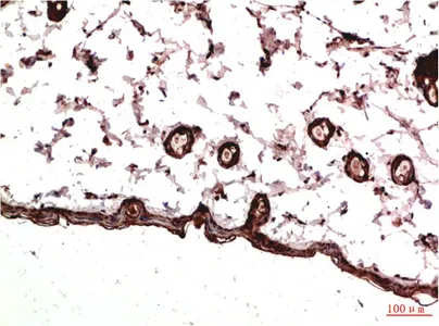
Immunohistochemical analysis of paraffin-embedded Rat Skin Tissue using PDGFR a Mouse mAb diluted at 1:200.

No documents available.