
PerCP/Cyanine5.5 Anti-Mouse CD8a Antibody[53-6.7]
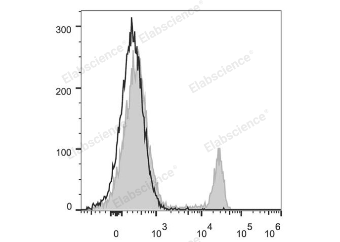
C57BL/6 murine splenocytes are stained with PerCP/Cyanine5.5 Anti-Mouse CD8a Antibody (filled gray histogram). Unstained splenocytes (empty black histogram) are used as control.
| Catalog Number: | MAB1104UJ |
| Conjugate: | PerCP/Cyanine5.5 is designed to be excited by the blue laser (488 nm) and detected using an optical filter centered near 675 nm (e.g., a 690/50 nm bandpass filter). |
| Size: | 100μg |
| Concentration: | 0.2 mg/mL |
| Host: | Rat |
| Isotype: | Rat IgG2a, κ |
| Clone: | 53-6.7 |
| Reactivity: | Mouse |
| Applications: | FCM - Each lot of this antibody is quality control tested by flow cytometric analysis. Please check your vial before the experiment. Since applications vary, the appropriate dilutions must be determined for individual use. We suggest each investigator should titrate the reagent to obtain optimal results [The recommended concentration is 0.1-1 μg/10<sup>6</sup> cells in 100 μL volume]. |
| Form: | Liquid |
CD8, also known as Lyt-2, Ly-2, or T8, consists of disulfide-linked α and β chains that form the α(CD8a)/β(CD8b) heterodimer and α/α homodimer. CD8a is a 34 kD protein that belongs to the immunoglobulin family. The CD8 α/β heterodimer is expressed on the surface of most thymocytes and a subset of mature TCR α/β T cells. CD8 expression on mature T cells is non-overlapping with CD4. The CD8 α/α homodimer is expressed on a subset of γ/δ TCR-bearing T cells, NK cells, intestinal intraepithelial lymphocytes, and lymphoid dendritic cells. CD8 is an antigen co-receptor on T cells that interacts with MHC class I on antigen-presenting cells or epithelial cells. CD8 promotes T cell activation through its association with the TCR complex and protein tyrosine kinase lck.
Applications
FCM - Each lot of this antibody is quality control tested by flow cytometric analysis. Please check your vial before the experiment. Since applications vary, the appropriate dilutions must be determined for individual use. We suggest each investigator should titrate the reagent to obtain optimal results [The recommended concentration is 0.1-1 μg/10<sup>6</sup> cells in 100 μL volume].