Phospho-AKT-S473 Rabbit mAb

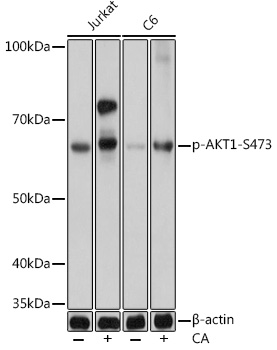
Western blot analysis of various lysates using Phospho-AKT1-S473 mAb  at 1:1000 dilution. Both Jurkat cells and C6 cells were treated by Calyculin A  at 37℃ for 30 minutes.
Secondary antibody: HRP-conjugated Goat anti-Rabbit IgG   at 1:10000 dilution.
Lysates/proteins: 25μg per lane.
Blocking buffer: 3% nonfat dry milk in TBST.
Detection: ECL Basic Kit .
Exposure time: 1s.

Western blot analysis of various lysates using Phospho-AKT1-S473 mAb at 1:1000 dilution. Both Jurkat cells and C6 cells were treated by Calyculin A at 37℃ for 30 minutes. Secondary antibody: HRP-conjugated Goat anti-Rabbit IgG at 1:10000 dilution. Lysates/proteins: 25μg per lane. Blocking buffer: 3% nonfat dry milk in TBST. Detection: ECL Basic Kit . Exposure time: 1s.

Catalog Number:AB0637
Conjugate:Unconjugated
Size:100 ug
Concentration:1mg/ml
Host:Rabbit
Isotype:IgG
Clone:ARC0169
Immunogen:A synthetic phosphorylated peptide around S473 of human Akt1 (P31749).
Reactivity:Human,Mouse,Rat
Applications:WB,1:500 - 1:2000 IHC-P,1:50 - 1:200
Molecular Weight:60kDa
Purification:Affinity purification
Form:liquid

This gene encodes one of the three members of the human AKT serine-threonine protein kinase family which are often referred to as protein kinase B alpha, beta, and gamma. These highly similar AKT proteins all have an N-terminal pleckstrin homology domain, a serine/threonine-specific kinase domain and a C-terminal regulatory domain. These proteins are phosphorylated by phosphoinositide 3-kinase (PI3K). AKT/PI3K forms a key component of many signalling pathways that involve the binding of membrane-bound ligands such as receptor tyrosine kinases, G-protein coupled receptors, and integrin-linked kinase. These AKT proteins therefore regulate a wide variety of cellular functions including cell proliferation, survival, metabolism, and angiogenesis in both normal and malignant cells. AKT proteins are recruited to the cell membrane by phosphatidylinositol 3,4,5-trisphosphate (PIP3) after phosphorylation of phosphatidylinositol 4,5-bisphosphate (PIP2) by PI3K. Subsequent phosphorylation of both threonine residue 308 and serine residue 473 is required for full activation of the AKT1 protein encoded by this gene. Phosphorylation of additional residues also occurs, for example, in response to insulin growth factor-1 and epidermal growth factor. Protein phosphatases act as negative regulators of AKT proteins by dephosphorylating AKT or PIP3. The PI3K/AKT signalling pathway is crucial for tumor cell survival. Survival factors can suppress apoptosis in a transcription-independent manner by activating AKT1 which then phosphorylates and inactivates components of the apoptotic machinery. AKT proteins also participate in the mammalian target of rapamycin (mTOR) signalling pathway which controls the assembly of the eukaryotic translation initiation factor 4F (eIF4E) complex and this pathway, in addition to responding to extracellular signals from growth factors and cytokines, is disregulated in many cancers. Mutations in this gene are associated with multiple types of cancer and excessive tissue growth including Proteus syndrome and Cowden syndrome 6, and breast, colorectal, and ovarian cancers. Multiple alternatively spliced transcript variants have been found for this gene.

Applications

WB,1:500 - 1:2000 IHC-P,1:50 - 1:200

Immunogen

A synthetic phosphorylated peptide around S473 of human Akt1 (P31749).
Target Background

This gene encodes one of the three members of the human AKT serine-threonine protein kinase family which are often referred to as protein kinase B alpha, beta, and gamma. These highly similar AKT proteins all have an N-terminal pleckstrin homology domain, a serine/threonine-specific kinase domain and a C-terminal regulatory domain. These proteins are phosphorylated by phosphoinositide 3-kinase (PI3K). AKT/PI3K forms a key component of many signalling pathways that involve the binding of membrane-bound ligands such as receptor tyrosine kinases, G-protein coupled receptors, and integrin-linked kinase. These AKT proteins therefore regulate a wide variety of cellular functions including cell proliferation, survival, metabolism, and angiogenesis in both normal and malignant cells. AKT proteins are recruited to the cell membrane by phosphatidylinositol 3,4,5-trisphosphate (PIP3) after phosphorylation of phosphatidylinositol 4,5-bisphosphate (PIP2) by PI3K. Subsequent phosphorylation of both threonine residue 308 and serine residue 473 is required for full activation of the AKT1 protein encoded by this gene. Phosphorylation of additional residues also occurs, for example, in response to insulin growth factor-1 and epidermal growth factor. Protein phosphatases act as negative regulators of AKT proteins by dephosphorylating AKT or PIP3. The PI3K/AKT signalling pathway is crucial for tumor cell survival. Survival factors can suppress apoptosis in a transcription-independent manner by activating AKT1 which then phosphorylates and inactivates components of the apoptotic machinery. AKT proteins also participate in the mammalian target of rapamycin (mTOR) signalling pathway which controls the assembly of the eukaryotic translation initiation factor 4F (eIF4E) complex and this pathway, in addition to responding to extracellular signals from growth factors and cytokines, is disregulated in many cancers. Mutations in this gene are associated with multiple types of cancer and excessive tissue growth including Proteus syndrome and Cowden syndrome 6, and breast, colorectal, and ovarian cancers. Multiple alternatively spliced transcript variants have been found for this gene.

Storage

Store at -20℃. Avoid freeze / thaw cycles.

Buffer

PBS with 0.02% sodium azide,0.05% BSA,50% glycerol,pH7.3.

Western blot analysis of various lysates using Phospho-AKT1-S473 mAb at 1:1000 dilution. Both Jurkat cells and C6 cells were treated by Calyculin A at 37℃ for 30 minutes. Secondary antibody: HRP-conjugated Goat anti-Rabbit IgG at 1:10000 dilution. Lysates/proteins: 25μg per lane. Blocking buffer: 3% nonfat dry milk in TBST. Detection: ECL Basic Kit . Exposure time: 1s.

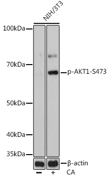
Western blot analysis of lysates from NIH/3T3 cells, using Phospho-AKT1-S473 mAb at 1:1000 dilution. NIH/3T3 cells were treated by Calyculin A at 37℃ for 30 minutes. Secondary antibody: HRP-conjugated Goat anti-Rabbit IgG at 1:10000 dilution. Lysates/proteins: 25μg per lane. Blocking buffer: 3% nonfat dry milk in TBST. Detection: ECL Basic Kit . Exposure time: 10s.

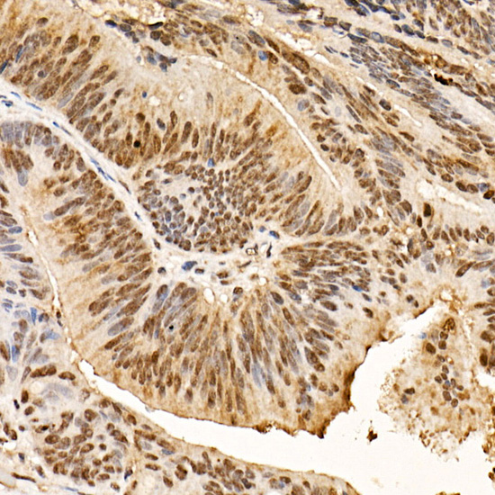
Immunohistochemistry analysis of paraffin-embedded Human colon carcinoma using Phospho-AKT1-S473 Rabbit mAb at dilution of 1:100 . High pressure antigen retrieval performed with 0.01M Citrate Bufferr prior to IHC staining.

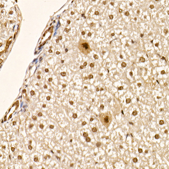
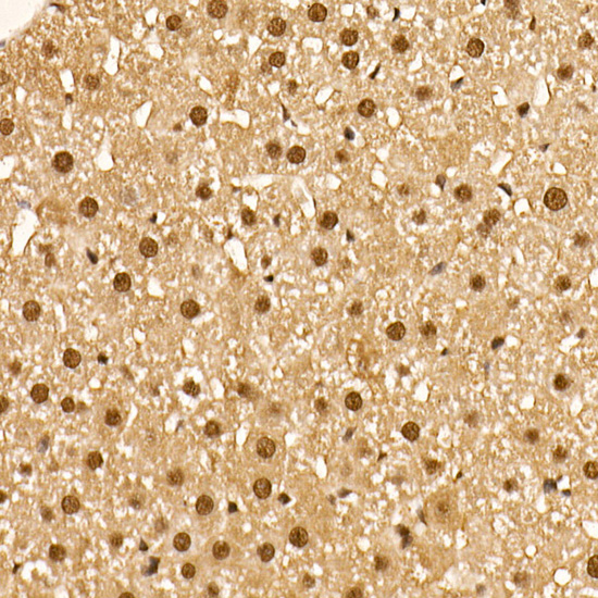
Immunohistochemistry analysis of paraffin-embedded Mouse liver using Phospho-AKT1-S473 Rabbit mAb at dilution of 1:100 . High pressure antigen retrieval performed with 0.01M Citrate Bufferr prior to IHC staining.

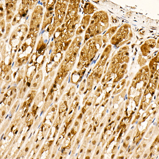
Immunohistochemistry analysis of paraffin-embedded Mouse stomach using Phospho-AKT1-S473 Rabbit mAb at dilution of 1:100 . High pressure antigen retrieval performed with 0.01M Citrate Bufferr prior to IHC staining.

Immunohistochemistry analysis of paraffin-embedded Rat liver using Phospho-AKT1-S473 Rabbit mAb at dilution of 1:100 . High pressure antigen retrieval performed with 0.01M Citrate Bufferr prior to IHC staining.

No documents available.