
Phospho-AKT1 (S473) Rabbit Monoclonal Antibody
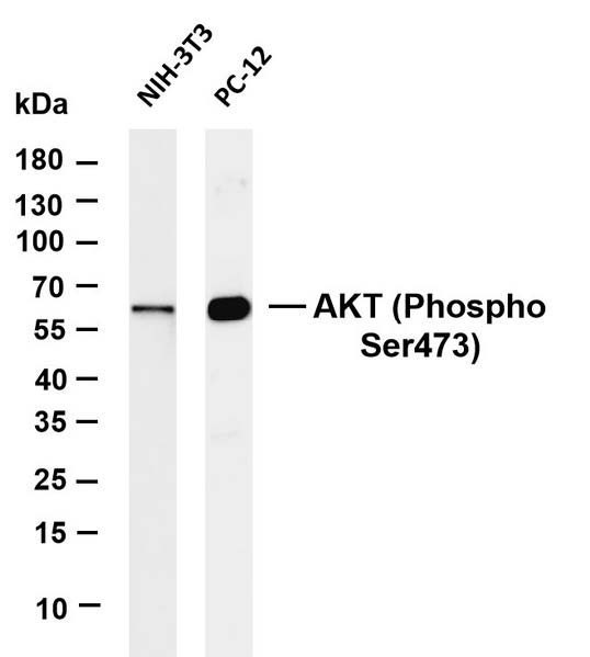
Various whole cell lysates were
separated by 4-20% SDS-PAGE, and
the membrane was blotted with antiAKT (Phospho Ser473) antibody.
The HRP-conjugated Goat anti-Rabbit
IgG (H + L) antibody was used to detect
the antibody.
Lane 1: NIH-3T3
Lane 2: PC-12
Observed band size: 60kDa

Various whole cell lysates were separated by 4-20% SDS-PAGE, and the membrane was blotted with antiAKT (Phospho Ser473) antibody. The HRP-conjugated Goat anti-Rabbit IgG (H + L) antibody was used to detect the antibody. Lane 1: NIH-3T3 Lane 2: PC-12 Observed band size: 60kDa

Catalog Number:MAB-94111
Conjugate:Unconjugated
Size:100 ug
Concentration:1mg/ml
Host:Rabbit
Isotype:IgG
Clone:D9E
Immunogen:The antiserum was produced against synthesized peptide derived from human Akt around the phosphorylation site of Ser473. AA range:431-480
Reactivity:Human;Mouse;Rat;Chicken
Applications:Western Blot: 1/500 - 1/2000Immunofluorescence: 1:50-200 Immunohistochemistry: 1/100 - 1/300
Molecular Weight:60 kDa
Purification:The antibody was affinity-purified from rabbit antiserum by affinity-chromatography using epitope-specific immunogen.
Form:liquid
Synonyms:AKT;CWS6;PKB;PKB-ALPHA;PRKBA;RAC;RAC-ALPHA;AKT1

Applications

Western Blot: 1/500 - 1/2000Immunofluorescence: 1:50-200 Immunohistochemistry: 1/100 - 1/300

Immunogen

The antiserum was produced against synthesized peptide derived from human Akt around the phosphorylation site of Ser473. AA range:431-480

Target Background

Background: Akt, also referred to as PKB or Rac, plays a critical role in controlling survival and apoptosis (1-3). This protein kinase is activated by insulin and various growth and survival factors to function in a wortmannin-sensitive pathway involving PI3 kinase (2,3). Akt is activated by phospholipid binding and activation loop phosphorylation at Thr308 by PDK1 (4) and by phosphorylation within the carboxy terminus at Ser473. The previously elusive PDK2 responsible for phosphorylation of Akt at Ser473 has been identified as mammalian target of rapamycin (mTOR) in a rapamycin-insensitive complex with rictor and Sin1 (5,6). Akt promotes cell survival by inhibiting apoptosis through phosphorylation and inactivation of several targets, including Bad (7), forkhead transcription factors (8), c-Raf (9), and caspase-9. PTEN phosphatase is a major negative regulator of the PI3 kinase/Akt signaling pathway (10). LY294002 is a specific PI3 kinase inhibitor (11). Another essential Akt function is the regulation of glycogen synthesis through phosphorylation and inactivation of GSK-3 and (12,13). Akt may also play a role in insulin stimulation of glucose transport (12). In addition to its role in survival and glycogen synthesis, Akt is involved in cell cycle regulation by preventing GSK-3 -mediated phosphorylation and degradation of cyclin D1 (14) and by negatively regulating the cyclin dependent kinase inhibitors p27 Kip (15) and p21 Waf1/CIP1 (16). Akt also plays a critical role in cell growth by directly phosphorylating mTOR in a rapamycin-sensitive complex containing raptor (17). More importantly, Akt phosphorylates and inactivates tuberin (TSC2), an inhibitor of mTOR within the mTOR-raptor complex (18,19). Specificity/Sensitivity: Phospho-Akt1 (Ser473) (D7F10) Rabbit mAb (Akt1 Specific) recognizes endogenous levels of Akt1 protein only when phosphorylated at Ser473. It does not detect Akt2 protein when phosphorylated at Ser474.

Synonyms:AKT;CWS6;PKB;PKB-ALPHA;PRKBA;RAC;RAC-ALPHA;AKT1

Storage

Store at -20℃. Avoid repeated freeze-thaw cycles.

Buffer

Liquid in PBS containing 50% glycerol, 0.5% BSA and 0.02% sodium azide.

Various whole cell lysates were separated by 4-20% SDS-PAGE, and the membrane was blotted with antiAKT (Phospho Ser473) antibody. The HRP-conjugated Goat anti-Rabbit IgG (H + L) antibody was used to detect the antibody. Lane 1: NIH-3T3 Lane 2: PC-12 Observed band size: 60kDa

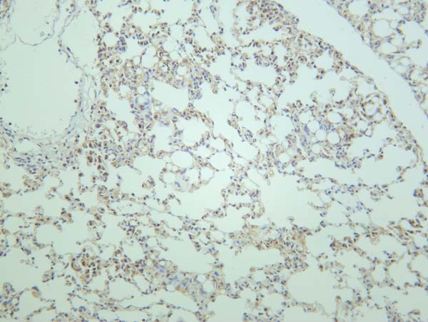
Mouse lung was stained with antiAKT (Phospho Ser473) rabbit antibod

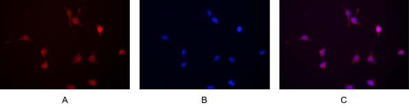
Immunofluorescence analysis of HEK293. Picture A: AKT antibody (red). Picture B: DAPI (blue). Picture C: Merge of A+B

No documents available.