Phospho-AMPKα1/2 (T183/172)

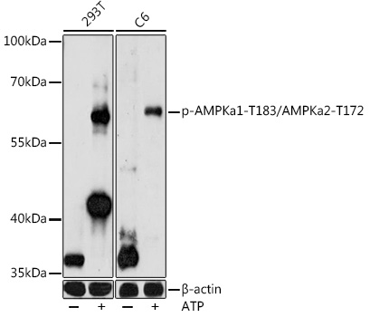
Western blot analysis of extracts of various cell lines, using Phospho-AMPKa1-T183/AMPKa2-T172 antibody at 1:1000 dilution.293T and C6 cells were treated by ATP(5 mM) at 30℃ for 1 hour. Secondary antibody: HRP Goat Anti-Rabbit IgG (H+L) at 1:10000 dilution. Lysates/proteins: 25ug per lane. Blocking buffer: 3% nonfat dry milk in TBST. Detection: ECL West Pico Plus.   Exposure time: 90s.

Western blot analysis of extracts of various cell lines, using Phospho-AMPKa1-T183/AMPKa2-T172 antibody at 1:1000 dilution.293T and C6 cells were treated by ATP(5 mM) at 30℃ for 1 hour. Secondary antibody: HRP Goat Anti-Rabbit IgG (H+L) at 1:10000 dilution. Lysates/proteins: 25ug per lane. Blocking buffer: 3% nonfat dry milk in TBST. Detection: ECL West Pico Plus. Exposure time: 90s.

Catalog Number:ABP-0883
Conjugate:Unconjugated
Size:100 ug
Concentration:1mg/ml
Host:Rb
Isotype:IgG
Clone:Poly
Immunogen:A phospho specific peptide corresponding to residues surrounding T172 of human AMPKα
Reactivity:Hu,Rt
Applications:WB
Molecular Weight:64kDa
Purification:Affinity purification
Form:liquid
Synonyms:AMPKa1/AMPKa2

Applications

WB

Immunogen

A phospho specific peptide corresponding to residues surrounding T172 of human AMPKα

Target Background

The protein encoded by this gene belongs to the ser/thr protein kinase family. It is the catalytic subunit of the 5'-prime-AMP-activated protein kinase (AMPK). AMPK is a cellular energy sensor conserved in all eukaryotic cells. The kinase activity of AMPK is activated by the stimuli that increase the cellular AMP/ATP ratio. AMPK regulates the activities of a number of key metabolic enzymes through phosphorylation. It protects cells from stresses that cause ATP depletion by switching off ATP-consuming biosynthetic pathways. Alternatively spliced transcript variants encoding distinct isoforms have been observed.

Synonyms:AMPKa1/AMPKa2

Storage

Store at -20℃. Avoid freeze / thaw cycles

Buffer

PBS with 0.02% sodium azide, 50% glycerol, pH7.3.

Western blot analysis of extracts of various cell lines, using Phospho-AMPKa1-T183/AMPKa2-T172 antibody at 1:1000 dilution.293T and C6 cells were treated by ATP(5 mM) at 30℃ for 1 hour. Secondary antibody: HRP Goat Anti-Rabbit IgG (H+L) at 1:10000 dilution. Lysates/proteins: 25ug per lane. Blocking buffer: 3% nonfat dry milk in TBST. Detection: ECL West Pico Plus. Exposure time: 90s.

No documents available.