Phospho-ERK1/ERK2 (p44/p42 MAPK) (T202/Y204)
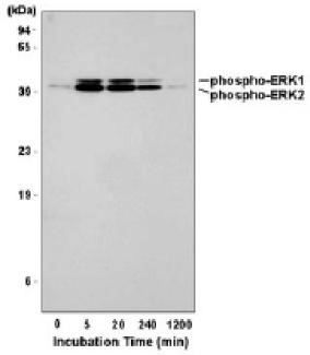
Detection of ERK1 and ERK2 phosphorylated at T202/Y204 and T185/Y187, respectively. Human HeLa cells were incubated with 200 nM PMA for the indicated times. Total cell lysates in gel sample buffer were resolved by SDS-PAGE.

Detection of ERK1 and ERK2 phosphorylated at T202/Y204 and T185/Y187, respectively. Human HeLa cells were incubated with 200 nM PMA for the indicated times. Total cell lysates in gel sample buffer were resolved by SDS-PAGE.

Catalog Number:ABP-83922
Conjugate:Unconjugated
Size:100 ug
Concentration:1mg/ml
Host:Rabbit
Isotype:IgG
Clone:Poly
Immunogen:Peptide derived from the protein area including conserved pT-E-pY motif of activated Erk 1,2
Reactivity:Hu, Ms, Rt
Applications:Western blotting:1:1000 Immunohistochemistry:1:100-500 Immunofluorescence: 1:100-500 Immunocytochemistry: 1:100
Molecular Weight:42,44kDa
Form:liquid

Applications

Western blotting:1:1000 Immunohistochemistry:1:100-500 Immunofluorescence: 1:100-500 Immunocytochemistry: 1:100

Immunogen

Peptide derived from the protein area including conserved pT-E-pY motif of activated Erk 1,2
Target Background

The protein encoded by this gene is a member of the MAP kinase family. MAP kinases, also known as extracellular signal-regulated kinases (ERKs), act in a signaling cascade that regulates various cellular processes such as proliferation, differentiation, and cell cycle progression in response to a variety of extracellular signals. This kinase is activated by upstream kinases, resulting in its translocation to the nucleus where it phosphorylates nuclear targets. Alternatively spliced transcript variants encoding different protein isoforms have been described.

Storage

The antibody can be stored at 2° - 8° C for 1 month without detectable loss of activity. Antibody can also be aliquotted and stored frozen at -20° C to -70° C in a manual defrost freezer for six months without detectable loss of activity. Avoid repeated freeze-thaw cycles.

Buffer

20 mM Tris Hcl, pH 8 10 mg/mL BSA - 0,05% Sodium Azide

Detection of ERK1 and ERK2 phosphorylated at T202/Y204 and T185/Y187, respectively. Human HeLa cells were incubated with 200 nM PMA for the indicated times. Total cell lysates in gel sample buffer were resolved by SDS-PAGE.

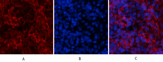
Immunofluorescence analysis of rat-lung tissue. 1,ERK 1/2 (phospho Thr202/Y204) Polyclonal Antibody(red) was diluted at 1:200(4°C,overnight). 2, Cy3 labled Secondary antibody was diluted at 1:300(room temperature, 50min).3, Picture B: DAPI(blue) 10min. Picture A:Target. Picture B: DAPI. Picture C: merge of A+B

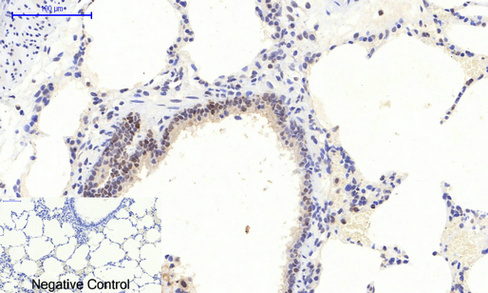
Immunohistochemical analysis of paraffin-embedded Rat-lung tissue. 1,ERK 1/2 (phospho Thr202/Y204) Polyclonal Antibody was diluted at 1:200(4°C,overnight). 2, Sodium citrate pH 6.0 was used for antibody retrieval(>98°C,20min). 3,Secondary antibody was diluted at 1:200(room tempeRature, 30min). Negative control was used by secondary antibody onl

No documents available.