Phospho-ERK1-T202/Y204 + ERK2-T185/Y187 Rabbit Monoclonal Antibody
Western blot analysis of extracts of various cell lines, using Phospho-ERK1-T202/Y204 + ERK2-
T185/Y187 Rabbit mAb at 1:1000 dilution.Both 293T cells and NIH/3T3 cells were treated by
ATP(5 mM) at 30℃ for 1 hour.
Secondary antibody: HRP Goat Anti-Rabbit IgG (H+L)at 1:10000 dilution.
Lysates/proteins: 25ug per lane.
Blocking buffer: 3% BSA.
Detection: ECL West Pico Plus.    
Exposure time: 10s.

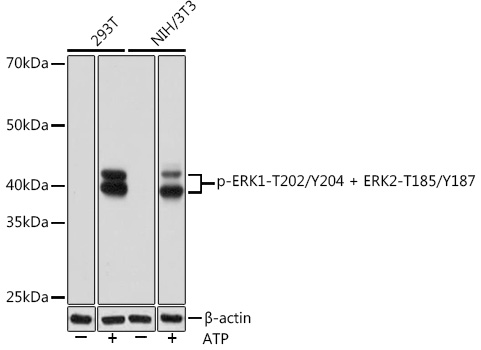
Western blot analysis of extracts of various cell lines, using Phospho-ERK1-T202/Y204 + ERK2- T185/Y187 Rabbit mAb at 1:1000 dilution.Both 293T cells and NIH/3T3 cells were treated by ATP(5 mM) at 30℃ for 1 hour. Secondary antibody: HRP Goat Anti-Rabbit IgG (H+L)at 1:10000 dilution. Lysates/proteins: 25ug per lane. Blocking buffer: 3% BSA. Detection: ECL West Pico Plus. Exposure time: 10s.

Catalog Number:MAB-94754
Conjugate:Unconjugated
Size:100 ug
Concentration:1mg/ml
Host:Rabbit
Isotype:IgG
Clone:ARC1595
Immunogen:A synthetic phosphorylated peptide around T202 & Y204 of human ERK1MK03 (
Reactivity:Human, Mouse, Rat
Applications:Western Blot: 1:500 - 1:2000 Immunohistochemistry: 1:50 - 1:200 Immunofluorescence: 1:50 - 1:200 Immunocytochemistry: 1:50 - 1:200
Molecular Weight:42kDa/44kDa
Purification:Affinity purification
Form:Liquid
Synonyms:ERK-1; ERK1; ERT2; HS44KDAP; HUMKER1A; P44ERK1; P44MAPK; PRKM3; p44-ERK1; p44- MAPK

Applications

Western Blot: 1:500 - 1:2000 Immunohistochemistry: 1:50 - 1:200 Immunofluorescence: 1:50 - 1:200 Immunocytochemistry: 1:50 - 1:200

Immunogen

A synthetic phosphorylated peptide around T202 & Y204 of human ERK1MK03 (
Target Background

The protein encoded by this gene is a member of the MAP kinase family. MAP kinases, also known as extracellular signal-regulated kinases (ERKs), act in a signaling cascade that regulates various cellular processes such as proliferation, differentiation, and cell cycle progression in response to a variety of extracellular signals. This kinase is activated by upstream kinases, resulting in its translocation to the nucleus where it phosphorylates nuclear targets. Alternatively spliced transcript variants encoding different protein isoforms have been described.

Synonyms:ERK-1; ERK1; ERT2; HS44KDAP; HUMKER1A; P44ERK1; P44MAPK; PRKM3; p44-ERK1; p44- MAPK

Storage

Store at -20℃. Avoid freeze / thaw cycles.

Buffer

PBS with 0.05% proclin300,0.05% BSA,50% glycerol,pH7.3.

Western blot analysis of extracts of various cell lines, using Phospho-ERK1-T202/Y204 + ERK2- T185/Y187 Rabbit mAb at 1:1000 dilution.Both 293T cells and NIH/3T3 cells were treated by ATP(5 mM) at 30℃ for 1 hour. Secondary antibody: HRP Goat Anti-Rabbit IgG (H+L)at 1:10000 dilution. Lysates/proteins: 25ug per lane. Blocking buffer: 3% BSA. Detection: ECL West Pico Plus. Exposure time: 10s.

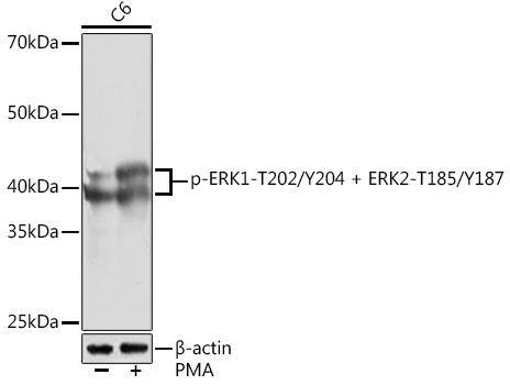
Western blot analysis of extracts of C6 cells, using Phospho-ERK1-T202/Y204 + ERK2-T185/Y187 Rabbit mAb at 1:1000 dilution.C6 cells were treated by PMA/TPA (200 nM) at 37℃ for 30 minutes after serum-starvation overnight. Secondary antibody: HRP Goat Anti-Rabbit IgG (H+L) at 1:10000 dilution. Lysates/proteins: 25ug per lane. Blocking buffer: 3% BSA. Detection: ECL West Pico Plus. Exposure time: 90s.

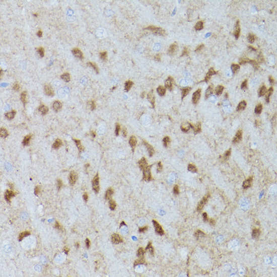
Immunohistochemistry of paraffinembedded rat brain using Phospho-ERK1- T202/Y204 + ERK2-T185/Y187 Rabbit mAb at dilution of 1:100 (40x lens).

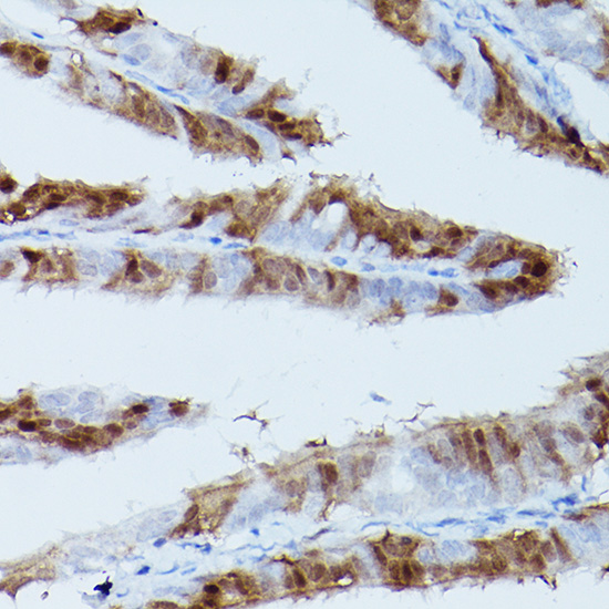
Immunohistochemistry of paraffinembedded rat fallopian tube using Phospho-ERK1-T202/Y204 + ERK2- T185/Y187 Rabbit mAb at dilution of 1:100 (40x lens).

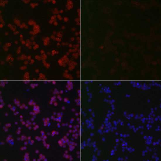
Immunofluorescence analysis of Jurkat treated with PMA and Jurkat using Phospho-ERK1-T202/Y204 + ERK2- T185/Y187 Rabbit mAb at dilution of 1:100 (40x lens). Blue: DAPI for nuclear staining.

No documents available.