Phospho Histone H2A.X (Ser139) Rabbit Polyclonal Antibody
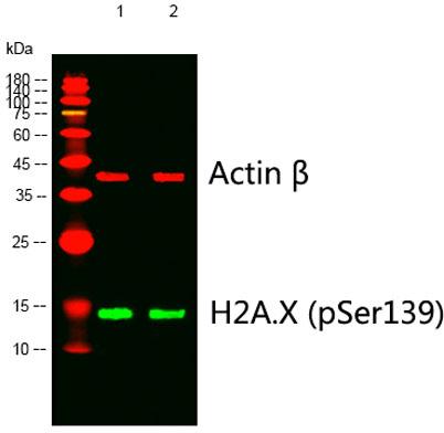
Western blot analysis of lysates from 1) 4T1, 2) 293 cells, (Green) primary antibody was diluted at 1:1000, 4°over
night, Dylight 800 secondary antibody was diluted at 1:10000, 37° 1hour. (Red) Actin β Monoclonal Antibody(5B7)

Western blot analysis of lysates from 1) 4T1, 2) 293 cells, (Green) primary antibody was diluted at 1:1000, 4°over night, Dylight 800 secondary antibody was diluted at 1:10000, 37° 1hour. (Red) Actin β Monoclonal Antibody(5B7)

Catalog Number:ABP-0640
Conjugate:Unconjugated
Size:100 ug
Concentration:1mg/ml
Host:Rabbit
Isotype:IgG
Clone:POLY
Immunogen:The antiserum was produced against synthesized peptide derived from human Histone H2A.X around the phosphorylation site of Ser139. AA range:94-143.
Reactivity:Human;Mouse;Rat;Hamster
Applications:Western Blot: 1:500 - 1:2000 Immunohistochemistry: 1:100 - 1:300 Immunofluorescence: 1:50-200 ELISA: 1:10000
Molecular Weight:15 kDa
Purification:The antibody was affinity-purified from rabbit antiserum by affinity-chromatography using epitope-specific immunogen.
Form:Liquid
Synonyms:H2AFX; H2AX; Histone H2A.x; H2a/x

Applications

Western Blot: 1:500 - 1:2000 Immunohistochemistry: 1:100 - 1:300 Immunofluorescence: 1:50-200 ELISA: 1:10000

Immunogen

The antiserum was produced against synthesized peptide derived from human Histone H2A.X around the phosphorylation site of Ser139. AA range:94-143.

Target Background

Histones are basic nuclear proteins that are responsible for the nucleosome structure of the chromosomal fiber in eukaryotes. Two molecules of each of the four core histones (H2A, H2B, H3, and H4) form an octamer, around which approximately 146 bp of DNA is wrapped in repeating units, called nucleosomes. The linker histone, H1, interacts with linker DNA between nucleosomes and functions in the compaction of chromatin into higHer order structures. This gene encodes a replication-independent histone that is a member of the histone H2A family, and generates two transcripts through the use of the conserved stem-loop termination motif, and the polyA addition motif.

Synonyms:H2AFX; H2AX; Histone H2A.x; H2a/x

Storage

SStore at -20°C. Avoid repeated freeze-thaw cycles.

Buffer

Liquid in PBS containing 50% glycerol, 0.5% BSA and 0.02% sodium azide.

Western blot analysis of lysates from 1) 4T1, 2) 293 cells, (Green) primary antibody was diluted at 1:1000, 4°over night, Dylight 800 secondary antibody was diluted at 1:10000, 37° 1hour. (Red) Actin β Monoclonal Antibody(5B7)

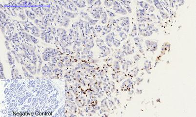
Immunohistochemical analysis of paraffin-embedded Human-stomach-cancer tissue. 1,Histone H2A.X (phospho Ser139) Polyclonal Antibody was diluted at 1:200(4°C,overnight). 2, Sodium citrate pH 6.0 was used for antibody retrieval(>98°C,20min). 3,Secondary antibody was diluted at 1:200(room tempeRature, 30min). Negative control was used by secondary antibody only.

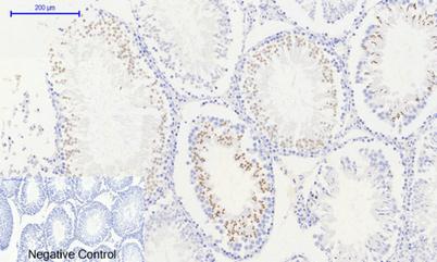
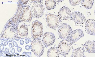
Immunohistochemical analysis of paraffin-embedded Mouse-testis tissue. 1,Histone H2A.X (phospho Ser139) Polyclonal Antibody was diluted at 1:200(4°C,overnight). 2, Sodium citrate pH 6.0 was used for antibody retrieval(>98°C,20min). 3,Secondary antibody was diluted at 1:200(room tempeRature, 30min). Negative control was used by secondary antibody only.

Immunohistochemical analysis of paraffin-embedded Rat-testis tissue. 1,Histone H2A.X (phospho Ser139) Polyclonal Antibody was diluted at 1:200(4°C,overnight). 2, Sodium citrate pH 6.0 was used for antibody retrieval(>98°C,20min). 3,Secondary antibody was

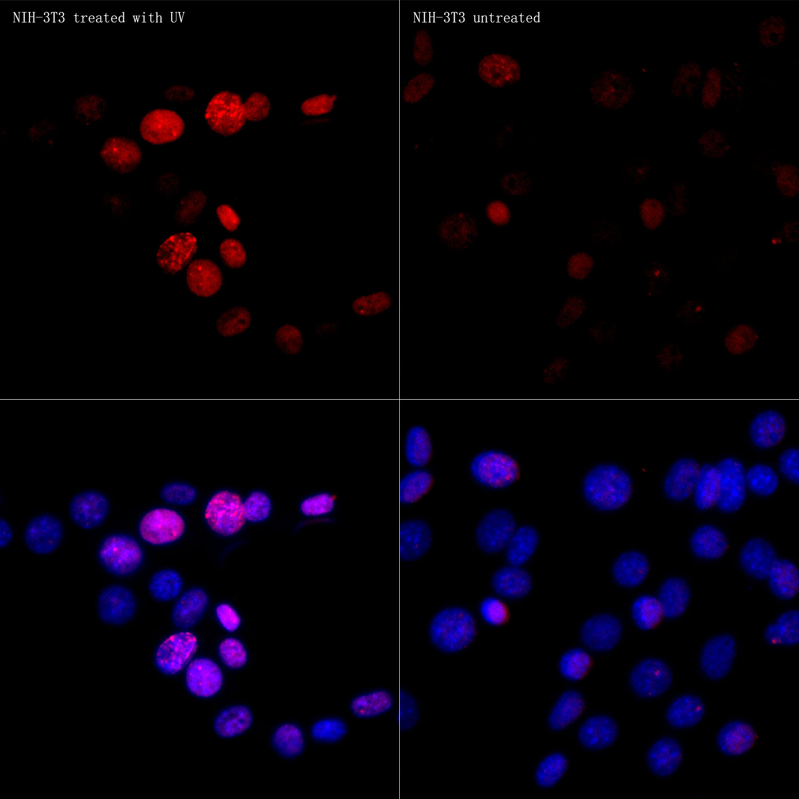
Immunofluorescence analysis of NIH-3T3 (treated with UV,30 mJ/cm2) and NIH/3T3 (untreated), using Phospho-Histone H2AXS139 Rabbit PAb at dilution of 1:50 (40x lens). Blue: DAPI for nuclear staining.

No documents available.