Phospho-Histone H2AFX-S139

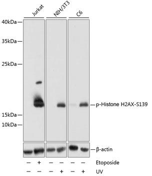
Western blot analysis of extracts of various cell lines, using Phospho-Histone H2AX-S139 antibody at 1:1000 dilution. Jurkat cells were treated by Etoposide (25 uM) for 5 hours. NIH/3T3 cells were treated by UV for 15-30 minutes. C6 cells were treated by UV for 15-30 minutes.  Secondary antibody: HRP Goat Anti-Rabbit IgG (H+L) at 1:10000 dilution. Lysates/proteins: 25ug per lane. Blocking buffer: 3% BSA. Detection: ECL West Pico Plus     Exposure time: 5s.

Western blot analysis of extracts of various cell lines, using Phospho-Histone H2AX-S139 antibody at 1:1000 dilution. Jurkat cells were treated by Etoposide (25 uM) for 5 hours. NIH/3T3 cells were treated by UV for 15-30 minutes. C6 cells were treated by UV for 15-30 minutes. Secondary antibody: HRP Goat Anti-Rabbit IgG (H+L) at 1:10000 dilution. Lysates/proteins: 25ug per lane. Blocking buffer: 3% BSA. Detection: ECL West Pico Plus Exposure time: 5s.

Catalog Number:ABP-0099
Conjugate:Unconjugated
Size:100 ug
Concentration:1mg/ml
Host:Rb
Isotype:IgG
Clone:Poly
Immunogen:A synthetic phosphorylated peptide around S139 of human Histone H2AX
Reactivity:Hu
Applications:Western Blot: 1:1000 Immunohistochemistry: 1:50 Immunofluorescence: 1:50
Molecular Weight:17kDa
Purification:Affinity purification
Form:liquid
Synonyms:H2AFX; H2A.X; H2A/X; H2AX; histone H2AX

Applications

Western Blot: 1:1000 Immunohistochemistry: 1:50 Immunofluorescence: 1:50

Immunogen

A synthetic phosphorylated peptide around S139 of human Histone H2AX
Target Background

Histones are basic nuclear proteins that are responsible for the nucleosome structure of the chromosomal fiber in eukaryotes. Two molecules of each of the four core histones (H2A, H2B, H3, and H4) form an octamer, around which approximately 146 bp of DNA is wrapped in repeating units, called nucleosomes. The linker histone, H1, interacts with linker DNA between nucleosomes and functions in the compaction of chromatin into higher order structures. This gene encodes a replication-independent histone that is a member of the histone H2A family, and generates two transcripts through the use of the conserved stemloop termination motif, and the polyA addition motif.

Synonyms:H2AFX; H2A.X; H2A/X; H2AX; histone H2AX

Storage

Store at -20℃. Avoid freeze / thaw cycles.

Buffer

PBS with 0.02% sodium azide, 50% glycerol, pH7.3.

Western blot analysis of extracts of various cell lines, using Phospho-Histone H2AX-S139 antibody at 1:1000 dilution. Jurkat cells were treated by Etoposide (25 uM) for 5 hours. NIH/3T3 cells were treated by UV for 15-30 minutes. C6 cells were treated by UV for 15-30 minutes. Secondary antibody: HRP Goat Anti-Rabbit IgG (H+L) at 1:10000 dilution. Lysates/proteins: 25ug per lane. Blocking buffer: 3% BSA. Detection: ECL West Pico Plus Exposure time: 5s.

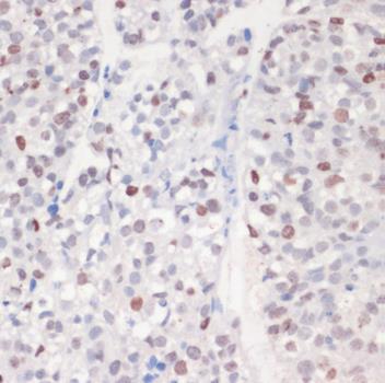
Immunohistochemistry of paraffin-embedded human gastric cancer using Phospho-Histone H2AX-S139 antibody at dilution of 1:200 (40x lens).

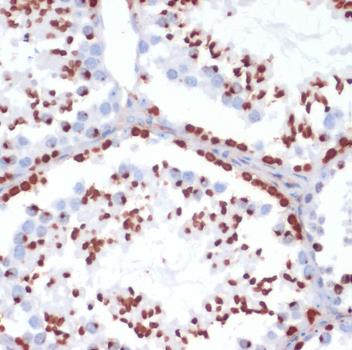
Immunohistochemistry of paraffin-embedded mouse testis using Phospho-Histone H2AX-S139 antibody at dilution of 1:200 (40x lens).

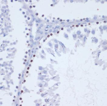
Immunohistochemistry of paraffin-embedded rat testis using Phospho-Histone H2AX-S139 antibody at dilution of 1:200 (40x lens).

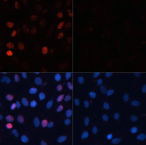
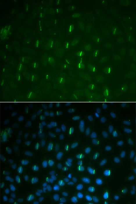
Immunofluorescence analysis of C6 cells using Phospho-Histone H2AX-S139 antibody at dilution of 1:100. Blue: DAPI for nuclear staining.C6 cells were treated by UV for 15-30 minutes at RT (left). Blue: DAPI for nuclear staining.

Immunofluorescence analysis of U2OS cells using Phospho-Histone H2AX-S139 antibody at dilution of 1:100. Blue: DAPI for nuclear staining.

No documents available.