Phospho-Histone H3-S10 Rabbit mAb

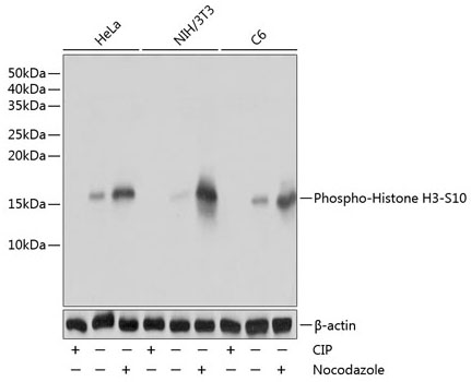
Western blot analysis of lysates from HeLa,NIH/3T3,C6 cells using Phospho-Histone H3- S10 Rabbit mAb  at 1:1000 dilution. HeLa,NIH/3T3 and C6 cells were treated with nocodazole (50 ng/mL) at 37℃ for 20 hours or treated with CIP(20uL/400ul) at 37℃ for 1 hour.
Secondary antibody: HRP-conjugated Goat anti-Rabbit IgG (H+L) ) at 1:10000 dilution.
Lysates/proteins: 25 μg per lane.
Blocking buffer: 3% nonfat dry milk in TBST.
Detection: ECL West Pico Plus.
Exposure time: 1s.

Western blot analysis of lysates from HeLa,NIH/3T3,C6 cells using Phospho-Histone H3- S10 Rabbit mAb at 1:1000 dilution. HeLa,NIH/3T3 and C6 cells were treated with nocodazole (50 ng/mL) at 37℃ for 20 hours or treated with CIP(20uL/400ul) at 37℃ for 1 hour. Secondary antibody: HRP-conjugated Goat anti-Rabbit IgG (H+L) ) at 1:10000 dilution. Lysates/proteins: 25 μg per lane. Blocking buffer: 3% nonfat dry milk in TBST. Detection: ECL West Pico Plus. Exposure time: 1s.

Catalog Number:MAB0002
Conjugate:Unconjugated
Size:100 ug
Concentration:1mg/ml
Host:Rabbit
Isotype:IgG
Clone:ARC0003
Immunogen:Synthetic peptide. This information is considered to be commercially sensitive
Reactivity:Human,Mouse, Rat, Other (Wide Range Predicted)
Applications:WB 1:500 - 1:1000 IHC-P 1:50 - 1:200 IF/ICC 1:50 - 1:200 ELISA Recommended starting concentration is 1 μg/mL. Please optimize the concentration based on your specific assay requirements.
Molecular Weight:17kDa
Purification:Affinity purification
Form:liquid
Synonyms:H3/A; H3C2; H3C3; H3C4; H3C6; H3C7; H3C8; H3FA; H3C10; H3C11; H3C12; HIST1H3A; Phospho-Histone H3-S10

Histones are basic nuclear proteins that are responsible for the nucleosome structure of the chromosomal fiber in eukaryotes. This structure consists of approximately 146 bp of DNA wrapped around a nucleosome, an octamer composed of pairs of each of the four core histones (H2A, H2B, H3, and H4). The chromatin fiber is further compacted through the interaction of a linker histone, H1, with the DNA between the nucleosomes to form higher order chromatin structures. This gene is intronless and encodes a replication-dependent histone that is a member of the histone H3 family. Transcripts from this gene lack polyA tails; instead, they contain a palindromic termination element. This gene is found in the large histone gene cluster on chromosome 6p22-p21.3.

Applications

WB 1:500 - 1:1000 IHC-P 1:50 - 1:200 IF/ICC 1:50 - 1:200 ELISA Recommended starting concentration is 1 μg/mL. Please optimize the concentration based on your specific assay requirements.

Immunogen

Synthetic peptide. This information is considered to be commercially sensitive

Target Background

Histones are basic nuclear proteins that are responsible for the nucleosome structure of the chromosomal fiber in eukaryotes. This structure consists of approximately 146 bp of DNA wrapped around a nucleosome, an octamer composed of pairs of each of the four core histones (H2A, H2B, H3, and H4). The chromatin fiber is further compacted through the interaction of a linker histone, H1, with the DNA between the nucleosomes to form higher order chromatin structures. This gene is intronless and encodes a replication-dependent histone that is a member of the histone H3 family. Transcripts from this gene lack polyA tails; instead, they contain a palindromic termination element. This gene is found in the large histone gene cluster on chromosome 6p22-p21.3.

Synonyms:H3/A; H3C2; H3C3; H3C4; H3C6; H3C7; H3C8; H3FA; H3C10; H3C11; H3C12; HIST1H3A; Phospho-Histone H3-S10

Storage

Store at -20 ℃ for one year. Avoid repeated freeze/thaw cycles.

Western blot analysis of lysates from HeLa,NIH/3T3,C6 cells using Phospho-Histone H3- S10 Rabbit mAb at 1:1000 dilution. HeLa,NIH/3T3 and C6 cells were treated with nocodazole (50 ng/mL) at 37℃ for 20 hours or treated with CIP(20uL/400ul) at 37℃ for 1 hour. Secondary antibody: HRP-conjugated Goat anti-Rabbit IgG (H+L) ) at 1:10000 dilution. Lysates/proteins: 25 μg per lane. Blocking buffer: 3% nonfat dry milk in TBST. Detection: ECL West Pico Plus. Exposure time: 1s.

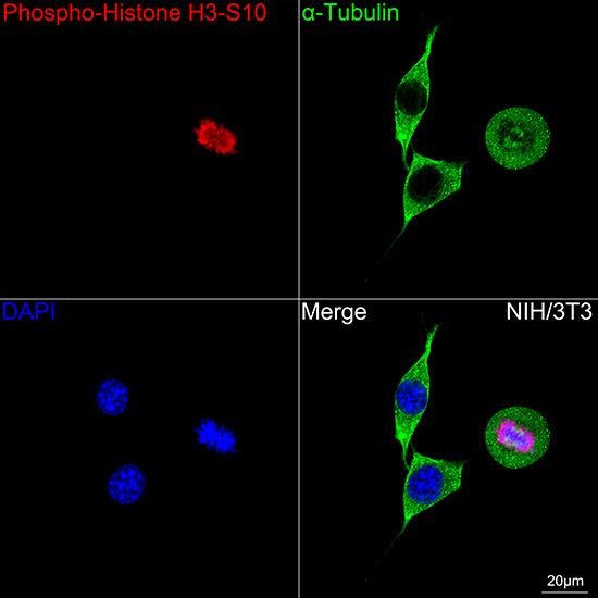
Confocal imaging of NIH/3T3 cells using Phospho-Histone H3-S10 Rabbit mAb (AP0002, dilution 1:100) followed by a further incubation withCy3 Goat Anti-Rabbit IgG (H+L) (, dilution 1:500) (Red). The cells were counterstained with α- Tubulin Mouse mAb ( dilution 1:400) followed by incubation with Alexa Fluro 488-conjugated Goat Anti- Mouse IgG (H+L) Ab (, dilution 1:500) (Green). DAPI was used for nuclear staining (Blue). Objective: 100x.
Immunohistochemistry analysis of paraffin-embedded Human oophoroma using Phospho-Histone H3-S10 Rabbit mAb at dilution of 1:100 (40x lens). High pressure antigen retrieval performed with 0.05M Tris/EDTA Buffer (pH 8.0) prior to IHC staining.

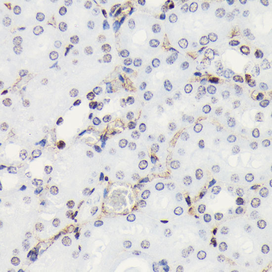
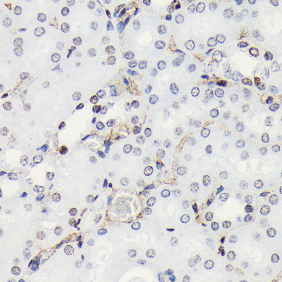
Immunohistochemistry analysis of paraffin-embedded Mouse kidney using Phospho-Histone H3-S10 Rabbit mAb at dilution of 1:100 (40x lens). High pressure antigen retrieval performed with 0.05M Tris/EDTA Buffer (pH 8.0) prior to IHC Staining

No documents available.