Phospho-IκBα-S32 Rabbit Monoclonal Antibody
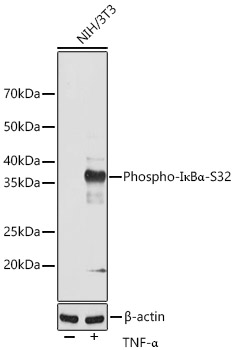
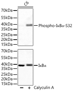
Western blot analysis of lysates from NIH/3T3 cells, using Phospho-IκBα-S32 Rabbit mAb (AP0707) at 1:1000 dilution. NIH/3T3 and C6 cells were treated with TNF-α (20 ng/mL) at 37℃ for 30 minutes. Secondary antibody: HRP-conjugated Goat anti-Rabbit IgG (H+L) at 1:10000 dilution. Lysates/proteins: 25μg per lane. Blocking buffer: 3% nonfat dry milk in TBST. Detection: ECL West Pico Plus. Exposure time: 120s.
| Catalog Number: | AB0707 |
| Conjugate: | Unconjugated |
| Size: | 100 ug |
| Concentration: | 1mg/ml |
| Host: | Rabbit |
| Isotype: | IgG |
| Clone: | ARC0147 |
| Immunogen: | Synthetic peptide. This information is considered to be commercially sensitive. |
| Reactivity: | Human,Mouse,Rat |
| Applications: | WB 1:500 - 1:2000 ELISA Recommended starting concentration is 1 μg/mL. Please optimize the concentration based on your specific assay requirements. |
| Molecular Weight: | 39kDa/ |
| Purification: | Affinity purification |
| Form: | liquid |
| Synonyms: | IKBA; MAD-3; NFKBI; EDAID2; Phospho-IκBα-S32 |
This gene encodes a member of the NF-kappa-B inhibitor family, which contain multiple ankrin repeat domains. The encoded protein interacts with REL dimers to inhibit NF-kappa-B/REL complexes which are involved in inflammatory responses. The encoded protein moves between the cytoplasm and the nucleus via a nuclear localization signal and CRM1-mediated nuclear export. Mutations in this gene have been found in ectodermal dysplasia anhidrotic with T-cell immunodeficiency autosomal dominant disease.
Applications
WB 1:500 - 1:2000 ELISA Recommended starting concentration is 1 μg/mL. Please optimize the concentration based on your specific assay requirements.
Immunogen
Synthetic peptide. This information is considered to be commercially sensitive.