Phospho-Jun-(S63)

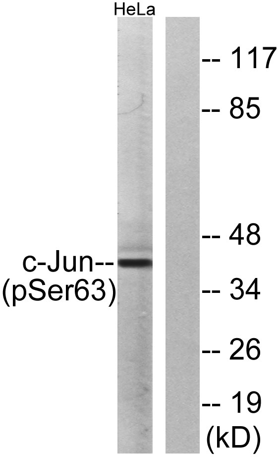
Western blot analysis of lysates from HeLa cells treated with UV, using c-Jun (Phospho-Ser63) Antibody. The lane on the right is blocked with the phospho peptide.

Western blot analysis of lysates from HeLa cells treated with UV, using c-Jun (Phospho-Ser63) Antibody. The lane on the right is blocked with the phospho peptide.

Catalog Number:ABP-0048
Conjugate:Unconjugated
Size:100 ug
Concentration:1mg/ml
Host:Rb
Isotype:IgG
Clone:Poly
Immunogen:A synthetic phosphorylated peptide around S63 of human AP-1.
Reactivity:Hu
Applications:Western Blot: 1:500 - 1:2000 Immunofluorescence: 1:50 - 1:200 Immunohistochemistry (paraffin-embedded tissues):1:100 - 1:300
Molecular Weight:48kDa
Purification:Affinity purification
Form:liquid
Synonyms:AP-1;AP1;c-Jun; JUN;

Applications

Western Blot: 1:500 - 1:2000 Immunofluorescence: 1:50 - 1:200 Immunohistochemistry (paraffin-embedded tissues):1:100 - 1:300

Immunogen

A synthetic phosphorylated peptide around S63 of human AP-1.
Target Background

This gene is the putative transforming gene of avian sarcoma virus 17. It encodes a protein which is highly similar to the viral protein, and which interacts directly with specific target DNA sequences to regulate gene expression. This gene is intronless and is mapped to 1p32-p31, a chromosomal region involved in both translocations and deletions in human malignancies.

Synonyms:AP-1;AP1;c-Jun; JUN;

Storage

Store at -20℃. Avoid freeze / thaw cycles.

Buffer

PBS with 0.02% sodium azide,50% glycerol,pH7.3.

Western blot analysis of lysates from HeLa cells treated with UV, using c-Jun (Phospho-Ser63) Antibody. The lane on the right is blocked with the phospho peptide.

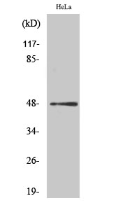
Western Blot analysis of various cells using Phospho-AP-1 (S63) Polyclonal Antibody diluted at 1:1000

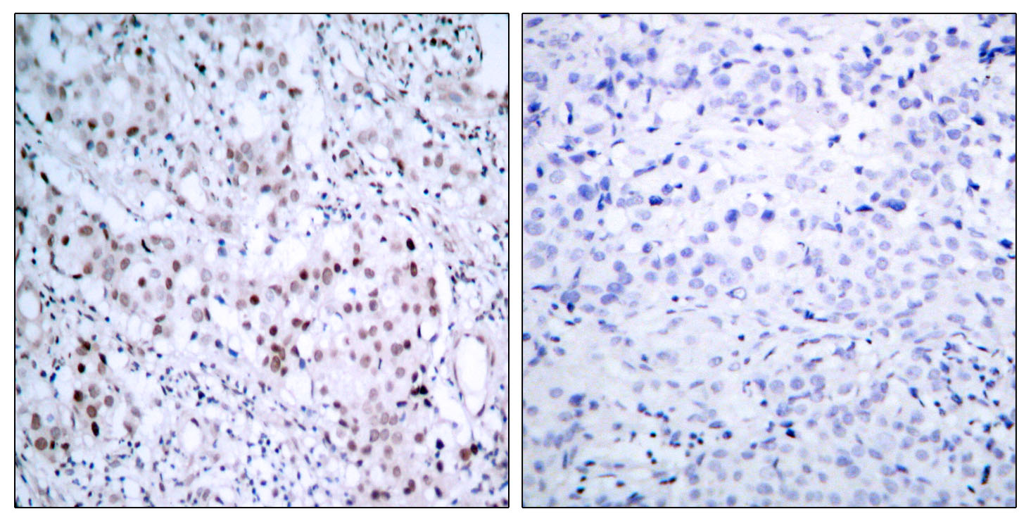
Immunohistochemistry analysis of paraffin-embedded human breast carcinoma, using c-Jun (Phospho-Ser63) Antibody. The picture on the right is blocked with the phospho peptide.

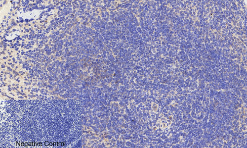
Immunohistochemical analysis of paraffin-embedded Mouse-brain tissue. 1,AP-1 (phospho Ser63) Polyclonal Antibody was diluted at 1:200(4°C,overnight). 2, Sodium citrate pH 6.0 was used for antibody retrieval(>98°C,20min). 3,Secondary antibody was diluted at 1:200(room tempeRature, 30min). Negative control was used by secondary antibody only.

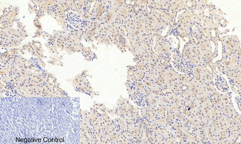
Immunohistochemical analysis of paraffin-embedded Rat-kidney tissue. 1,AP-1 (phospho Ser63) Polyclonal Antibody was diluted at 1:200(4°C,overnight). 2, Sodium citrate pH 6.0 was used for antibody retrieval(>98°C,20min). 3,Secondary antibody was diluted at 1:200(room tempeRature, 30min). Negative control was used by secondary antibody only.

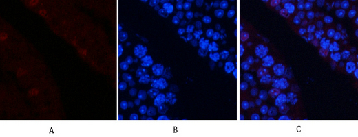
Immunofluorescence analysis of mouse-testis tissue. 1,AP-1 (phospho Ser63) Polyclonal Antibody(red) was diluted at 1:200(4°C,overnight). 2, Cy3 labled Secondary antibody was diluted at 1:300(room temperature, 50min).3, Picture B: DAPI(blue) 10min. Picture A:Target. Picture B: DAPI. Picture C: merge of A+B

No documents available.