
Phospho-Met (Y1349)

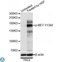
Western blot analysis of extracts of HeLa cells, using Phospho-MET-Y1349 antibody at 1:1000 dilution. HeLa cells were treated by HGF (40ng/ml) for 10 minutes after serum-starvation overnight. Secondary antibody: HRP Goat Anti-Rabbit IgG (H+L) at 1:10000 dilution. Lysates/proteins: 25ug per lane. Blocking buffer: 3% BSA. Detection: ECL Basic Kit. Exposure time: 90s.

Western blot analysis of extracts of HeLa cells, using Phospho-MET-Y1349 antibody at 1:1000 dilution. HeLa cells were treated by HGF (40ng/ml) for 10 minutes after serum-starvation overnight. Secondary antibody: HRP Goat Anti-Rabbit IgG (H+L) at 1:10000 dilution. Lysates/proteins: 25ug per lane. Blocking buffer: 3% BSA. Detection: ECL Basic Kit. Exposure time: 90s.

Catalog Number:ABP11206
Conjugate:Unconjugated
Size:100 ug
Concentration:1mg/ml
Host:Rb
Isotype:IgG
Clone:Poly
Immunogen:A phospho specific peptide corresponding to residues surrounding Y1349 of human MET
Reactivity:Hu
Applications:Western Blot: 1:500 - 1:2000 Immunohistochemistry 1:50 - 1:200 Immunofluorescence: 1:50 - 1:200
Molecular Weight:155.541 kDa
Purification:Affinity purification
Form:liquid

Applications

Western Blot: 1:500 - 1:2000 Immunohistochemistry 1:50 - 1:200 Immunofluorescence: 1:50 - 1:200

Immunogen

A phospho specific peptide corresponding to residues surrounding Y1349 of human MET
Target Background

This gene encodes a member of the receptor tyrosine kinase family of proteins and the product of the proto-oncogene MET. The encoded preproprotein is proteolytically processed to generate alpha and beta subunits that are linked via disulfide bonds to form the mature receptor. Further processing of the beta subunit results in the formation of the M10 peptide, which has been shown to reduce lung fibrosis. Binding of its ligand, hepatocyte growth factor, induces dimerization and activation of the receptor, which plays a role in cellular survival, embryogenesis, and cellular migration and invasion. Mutations in this gene are associated with papillary renal cell carcinoma, hepatocellular carcinoma, and various head and neck cancers. Amplification and overexpression of this gene are also associated with multiple

Storage

Store at -20°C, and avoid repeat freeze-thaw cycles

Buffer

PBS with 0.02% sodium azide, 50% glycerol, pH7.3

Western blot analysis of extracts of HeLa cells, using Phospho-MET-Y1349 antibody at 1:1000 dilution. HeLa cells were treated by HGF (40ng/ml) for 10 minutes after serum-starvation overnight. Secondary antibody: HRP Goat Anti-Rabbit IgG (H+L) at 1:10000 dilution. Lysates/proteins: 25ug per lane. Blocking buffer: 3% BSA. Detection: ECL Basic Kit. Exposure time: 90s.

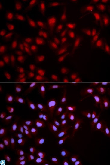
Immunofluorescence analysis of U2OS cells using Phospho-MET-Y1349 antibody Blue: DAPI for nuclear staining.

No documents available.