Phospho-NFKB-p65 (S536) Rabbit Polyclonal Antibody
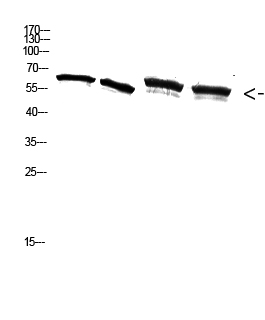
Western Blot analysis of A549 3T3 293T K562 cells using Antibody diluted at 2000. Secondary antibody was diluted at 1:20000

Western Blot analysis of A549 3T3 293T K562 cells using Antibody diluted at 2000. Secondary antibody was diluted at 1:20000

Catalog Number:ABP-0124
Conjugate:Unconjugated
Size:100 ug
Concentration:1mg/ml
Host:Rabbit
Isotype:IgG
Clone:Poly
Immunogen:The antiserum was produced against synthesized peptide derived from human NF-kappaB p65 around the phosphorylation site of Ser536. AA range:502-551
Reactivity:Hu, Ms, Rt, Monkey
Applications:Western Blot: 1/500 - 1/2000 Immunofluorescence: 1:50-200 Immunohistochemistry: 1/100 - 1/300. Immunoprecipitation: 2-5 ug/mg lysate
Molecular Weight:60kD
Purification:The antibody was affinity-purified from rabbit antiserum by affinity-chromatography using epitope-specific immunogen.
Form:liquid
Synonyms:RELA; NFKB3; Transcription factor p65; Nuclear factor NF-kappa-B p65 subunit; Nuclear factor of kappa light polypeptide gene enhancer in B-cells 3

Applications

Western Blot: 1/500 - 1/2000 Immunofluorescence: 1:50-200 Immunohistochemistry: 1/100 - 1/300. Immunoprecipitation: 2-5 ug/mg lysate

Immunogen

The antiserum was produced against synthesized peptide derived from human NF-kappaB p65 around the phosphorylation site of Ser536. AA range:502-551

Target Background

NF-kappa-B is a ubiquitous transcription factor involved in several biological processes. It is held in the cytoplasm in an inactive state by specific inhibitors. Upon degradation of the inhibitor, NF-kappa-B moves to the nucleus and activates transcription of specific genes. NF-kappa-B is composed of NFKB1 or NFKB2 bound to either REL, RELA, or RELB. The most abundant form of NF-kappa-B is NFKB1 complexed with the product of this gene, RELA. Four transcript variants encoding different isoforms have been found for this gene.

Synonyms:RELA; NFKB3; Transcription factor p65; Nuclear factor NF-kappa-B p65 subunit; Nuclear factor of kappa light polypeptide gene enhancer in B-cells 3

Storage

Store at -20℃. Avoid repeated freeze-thaw cycles.

Buffer

Liquid in PBS containing 50% glycerol, 0.5% BSA and 0.02% sodium azide.

Western Blot analysis of A549 3T3 293T K562 cells using Antibody diluted at 2000. Secondary antibody was diluted at 1:20000

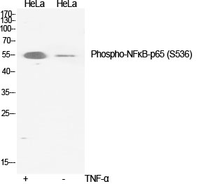
Western Blot analysis of various cells using Phospho-NFκB-p65 (S536) Polyclonal Antibody diluted at 1:2000

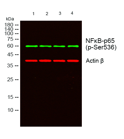
Western blot analysis of lysates from 1) A549, 2) 3T3, 3) 293T , 4)K562 cells, (Green) primary antibody was diluted at 1:1000, 4°over night, secondary antibody was diluted at 1:10000, 37° 1hour. (Red) Actin β Monoclonal Antibody(5B7) antibody was diluted at 1:5000 as loading control, 4° over night,secondary antibody was diluted at 1:10000, 37° 1hour.

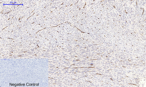
Immunohistochemical analysis of paraffin-embedded Human-uterus tissue. 1,NFκB-p65 (phospho Ser536) Polyclonal Antibody was diluted at 1:200(4°C,overnight). 2, Sodium citrate pH 6.0 was used for antibody retrieval(>98°C,20min). 3,Secondary antibody was diluted at 1:200(room tempeRature, 30min). Negative control was used by secondary antibody only.

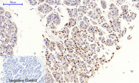
Immunohistochemical analysis of paraffin-embedded Human-stomach-cancer tissue. 1,NFκB-p65 (phospho Ser536) Polyclonal Antibody was diluted at 1:200(4°C,overnight). 2, Sodium citrate pH 6.0 was used for antibody retrieval(>98°C,20min). 3,Secondary antibody was diluted at 1:200(room tempeRature, 30min). Negative control was used by secondary antibody only.

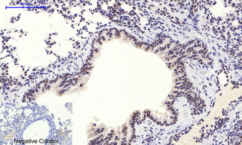
Immunohistochemical analysis of paraffin-embedded Mouse-lung tissue. 1,NFκB-p65 (phospho Ser536) Polyclonal Antibody was diluted at 1:200(4°C,overnight). 2, Sodium citrate pH 6.0 was used for antibody retrieval(>98°C,20min). 3,Secondary antibody was diluted at 1:200(room tempeRature, 30min). Negative control was used by secondary antibody only.

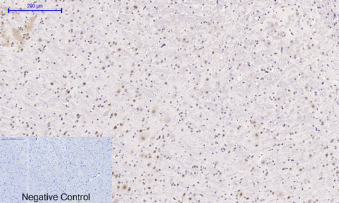
Immunohistochemical analysis of paraffin-embedded Mouse-brain tissue. 1,NFκB-p65 (phospho Ser536) Polyclonal Antibody was diluted at 1:200(4°C,overnight). 2, Sodium citrate pH 6.0 was used for antibody retrieval(>98°C,20min). 3,Secondary antibody was diluted at 1:200(room tempeRature, 30min). Negative control was used by secondary antibody only.

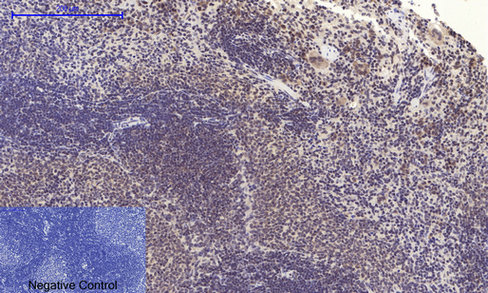
Immunohistochemical analysis of paraffin-embedded Rat-spleen tissue. 1,NFκB-p65 (phospho Ser536) Polyclonal Antibody was diluted at 1:200(4°C,overnight). 2, Sodium citrate pH 6.0 was used for antibody retrieval(>98°C,20min). 3,Secondary antibody was diluted at 1:200(room tempeRature, 30min). Negative control was used by secondary antibody only.

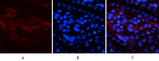
Immunofluorescence analysis of mouse-kidney tissue. 1,NFκB-p65 (phospho Ser536) Polyclonal Antibody(red) was diluted at 1:200(4°C,overnight). 2, Cy3 labled Secondary antibody was diluted at 1:300(room temperature, 50min).3, Picture B: DAPI(blue) 10min. Picture A:Target. Picture B: DAPI. Picture C: merge of A+B

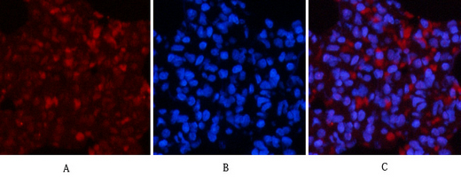
Immunofluorescence analysis of rat-lung tissue. 1,NFκB-p65 (phospho Ser536) Polyclonal Antibody(red) was diluted at 1:200(4°C,overnight). 2, Cy3 labled Secondary antibody was diluted at 1:300(room temperature, 50min).3, Picture B: DAPI(blue) 10min. Picture A:Target. Picture B: DAPI. Picture C: merge of A+B

No documents available.