Phospho-P70 S6K1 (T389) Rabbit Monoclonal Antibody
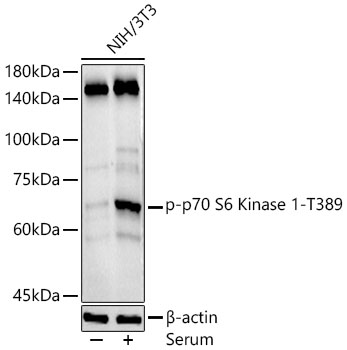
Western blot analysis of NIH/3T3, using Phospho-p70 S6K1 (T389) antibody at 1:500 dilution. NIH/3T3 cells were treated by 10% FBS at 37℃ for 30 minutes after serum-starvation overnight.
Secondary antibody: HRP Goat Anti-Rabbit IgG (H+L) at 1:10000 dilution.
Lysates/proteins: 25ug per lane.
Blocking buffer: 3% nonfat dry milk in TBST.
Detection: ECL Basic Kit 
Exposure time: 180s.

Western blot analysis of NIH/3T3, using Phospho-p70 S6K1 (T389) antibody at 1:500 dilution. NIH/3T3 cells were treated by 10% FBS at 37℃ for 30 minutes after serum-starvation overnight. Secondary antibody: HRP Goat Anti-Rabbit IgG (H+L) at 1:10000 dilution. Lysates/proteins: 25ug per lane. Blocking buffer: 3% nonfat dry milk in TBST. Detection: ECL Basic Kit Exposure time: 180s.

Catalog Number:MAB-94648
Conjugate:Unconjugated
Size:100 ug
Concentration:1mg/ml
Host:Rb
Isotype:IgG
Clone:108D2
Immunogen:A synthetic phosphorylated peptide around T389 of human P70 S6K
Reactivity:Hu, Ms, Rt
Applications:WB 1:500 - 1:2000 IF 1:50 - 1:200 ICC 1:50 - 1:200
Molecular Weight:70 kDa
Purification:Affinity purification
Form:liquid
Synonyms:PS6K;S6K;S6K-beta-1;S6K1;STK14A;p70 S6KA;p70(S6K)-alpha;p70-S6K;p70-alpha;P70 S6K;RPS6KB1;p70S6KA

Applications

WB 1:500 - 1:2000 IF 1:50 - 1:200 ICC 1:50 - 1:200

Immunogen

A synthetic phosphorylated peptide around T389 of human P70 S6K
Target Background

This gene encodes a member of the ribosomal S6 kinase family of serine/threonine kinases. The encoded protein responds to mTOR (mammalian target of rapamycin) signaling to promote protein synthesis, cell growth, and cell proliferation. Activity of this gene has been associated with human cancer. Alternatively spliced transcript variants have been observed. The use of alternative translation start sites results in isoforms with longer or shorter N-termini which may differ in their subcellular localizations. There are two pseudogenes for this gene on chromosome 17.

Synonyms:PS6K;S6K;S6K-beta-1;S6K1;STK14A;p70 S6KA;p70(S6K)-alpha;p70-S6K;p70-alpha;P70 S6K;RPS6KB1;p70S6KA

Storage

Store at -20℃. Avoid freeze / thaw cycles.

Buffer

PBS with 0.02% sodium azide,50% glycerol,pH7.3.

Western blot analysis of NIH/3T3, using Phospho-p70 S6K1 (T389) antibody at 1:500 dilution. NIH/3T3 cells were treated by 10% FBS at 37℃ for 30 minutes after serum-starvation overnight. Secondary antibody: HRP Goat Anti-Rabbit IgG (H+L) at 1:10000 dilution. Lysates/proteins: 25ug per lane. Blocking buffer: 3% nonfat dry milk in TBST. Detection: ECL Basic Kit Exposure time: 180s.

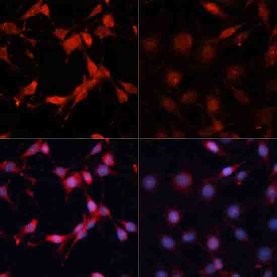
Immunofluorescence analysis of C6 cells using Phospho-p70 S6K1 (T389) antibody at dilution of 1:100. C6 cells were treated by Serum-starvation overnight at 37℃.

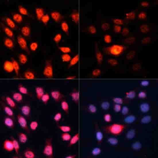
Immunofluorescence analysis of HeLa cells using Phospho-p70 S6K1 (T389) antibody at dilution of 1:100. HeLa cells were treated by Serum-starvation overnight at 37℃.

No documents available.