Phospho-Stat1 (Tyr701) Rabbit Polyclonal Antibody
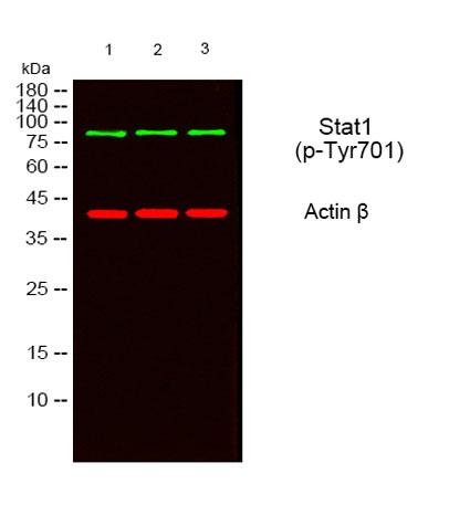
Western blot analysis of lysates from 1) MOUSE-BRAIN, 2) SY-SY5X ,3) COS7 cells, (Green)primary antibody was
diluted at 1:1000, 4°over night, secondary antibody(cat:RS23920)was diluted at 1:10000, 37° 1hour.
(Red) Actin β Monoclonal Antibody(5B7)  antibody was diluted at 1:5000 as loading control, 4° over
night,secondary antibody was diluted at 1:10000, 37° 1hour.

Western blot analysis of lysates from 1) MOUSE-BRAIN, 2) SY-SY5X ,3) COS7 cells, (Green)primary antibody was diluted at 1:1000, 4°over night, secondary antibody(cat:RS23920)was diluted at 1:10000, 37° 1hour. (Red) Actin β Monoclonal Antibody(5B7) antibody was diluted at 1:5000 as loading control, 4° over night,secondary antibody was diluted at 1:10000, 37° 1hour.

Catalog Number:ABP-0135
Conjugate:Unconjugated
Size:100 ug
Concentration:1mg/ml
Host:Rabbit
Isotype:IgG
Clone:Poly
Immunogen:The antiserum was produced against synthesized peptide derived from human STAT1 around the phosphorylation site of Tyr701. AA range:668-717.
Reactivity:Human;Mouse;Rat;Monkey
Applications:Western Blot: 1/500 - 1/2000 Immunofluorescence: 1:50-200 Immunohistochemistry: 1/100 - 1/300 Immunoprecipitation: 2-5 ug/mg lysate ELISA:1/10000
Molecular Weight:87 kDa
Purification:The antibody was affinity-purified from rabbit antiserum by affinity-chromatography using epitope-specific immunogen.
Form:liquid
Synonyms:STAT1; Signal transducer and activator of transcription 1-alpha/beta; Transcription factor ISGF-3 components p91/p84

Applications

Western Blot: 1/500 - 1/2000 Immunofluorescence: 1:50-200 Immunohistochemistry: 1/100 - 1/300 Immunoprecipitation: 2-5 ug/mg lysate ELISA:1/10000

Immunogen

The antiserum was produced against synthesized peptide derived from human STAT1 around the phosphorylation site of Tyr701. AA range:668-717.

Target Background

The protein encoded by this gene is a member of the STAT protein family. In response to cytokines and growth factors, STAT family members are phosphorylated by the receptor associated kinases, and then form homo- or heterodimers that translocate to the cell nucleus where they act as transcription activators. This protein can be activated by various ligands including interferon-alpha, interferon-gamma, EGF, PDGF and IL6. This protein mediates the expression of a variety of genes, which is thought to be important for cell viability in response to different cell stimuli and pathogens. Two alternatively spliced transcript variants encoding distinct isoforms have been described.

Synonyms:STAT1; Signal transducer and activator of transcription 1-alpha/beta; Transcription factor ISGF-3 components p91/p84

Storage

Store at -20°C. Avoid repeated freeze-thaw cycles.

Buffer

Liquid in PBS containing 50% glycerol, 0.5% BSA and 0.02% sodium azide

Western blot analysis of lysates from 1) MOUSE-BRAIN, 2) SY-SY5X ,3) COS7 cells, (Green)primary antibody was diluted at 1:1000, 4°over night, secondary antibody(cat:RS23920)was diluted at 1:10000, 37° 1hour. (Red) Actin β Monoclonal Antibody(5B7) antibody was diluted at 1:5000 as loading control, 4° over night,secondary antibody was diluted at 1:10000, 37° 1hour.

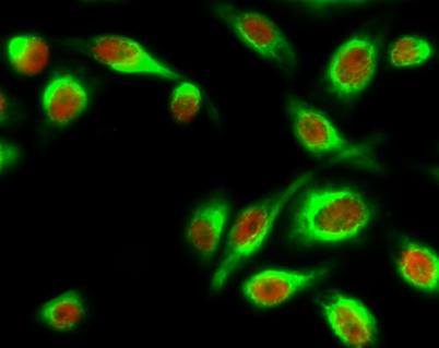
Immunofluorescence analysis of Hela cell. 1,Stat1 (phospho Tyr701) Polyclonal Antibody(red) was diluted at 1:200(4° overnight). HER2 Monoclonal Antibody(11H9)(green) was diluted at 1:200(4° overnight). 2, Goat Anti Rabbit Alexa Fluor 594

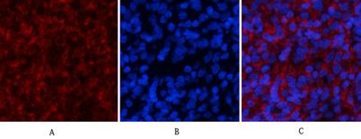
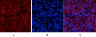
Immunofluorescence analysis of rat-spleen tissue. 1,Stat1 (phospho Tyr701) Polyclonal Antibody(red) was diluted at 1:200(4°C,overnight). 2, Cy3 labled Secondary antibody was diluted at 1:300(room temperature, 50min). 3, Picture B: DAPI(blue) 10min.

Immunofluorescence analysis of rat-spleen tissue. 1,Stat1 (phospho Tyr701) Polyclonal Antibody(red) was diluted at 1:200(4°C,overnight). 2, Cy3 labled Secondary antibody was diluted at 1:300(room temperature, 50min). 3, Picture B: DAPI(blue) 10min. Picture A:Target. Picture B: DAPI. Picture C: merge of A+B

No documents available.