
Phospho-VASP (S239)

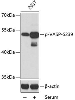
Western blot analysis of extracts of 293T cells, using Phospho-VASP-S239 antibody at 1:2000 dilution. 293T cells were treated by 10% FBS for 30 minutes after serum-starvation overnight. Secondary antibody: HRP Goat Anti- Rabbit IgG (H+L) at 1:10000 dilution. Lysates/proteins: 25ug per lane. Blocking buffer: 3% BSA. Detection: ECL West Pico Plus.    Exposure time: 90s.

Western blot analysis of extracts of 293T cells, using Phospho-VASP-S239 antibody at 1:2000 dilution. 293T cells were treated by 10% FBS for 30 minutes after serum-starvation overnight. Secondary antibody: HRP Goat Anti- Rabbit IgG (H+L) at 1:10000 dilution. Lysates/proteins: 25ug per lane. Blocking buffer: 3% BSA. Detection: ECL West Pico Plus. Exposure time: 90s.

Catalog Number:ABP-0187
Conjugate:Unconjugated
Size:100 ug
Concentration:1mg/ml
Host:Rb
Isotype:IgG
Clone:Poly
Immunogen:A phospho specific peptide corresponding to residues surrounding S239 of human VASP.
Reactivity:Hu
Applications:Western Blot: 1:500 - 1:1000
Molecular Weight:50kDa
Purification:Affinity purification
Form:liquid

Applications

Western Blot: 1:500 - 1:1000

Immunogen

A phospho specific peptide corresponding to residues surrounding S239 of human VASP.

Target Background

Vasodilator-stimulated phosphoprotein (VASP) is a member of the Ena-VASP protein family. Ena-VASP family members contain an EHV1 N-terminal domain that binds proteins containing E/DFPPPPXD/E motifs and targets Ena-VASP proteins to focal adhesions. In the mid-region of the protein, family members have a proline-rich domain that binds SH3 and WW domain-containing proteins. Their C-terminal EVH2 domain mediates tetramerization and binds both G and F actin. VASP is associated with filamentous actin formation and likely plays a widespread role in cell adhesion and motility. VASP may also be involved in the intracellular signaling pathways that regulate integrin-extracellular matrix interactions. VASP is regulated by the cyclic nucleotide-dependent kinases PKA and PKG.

Storage

Store at -20℃. Avoid freeze / thaw cycles

Buffer

PBS with 0.02% sodium azide,50% glycerol,pH7.3.

Western blot analysis of extracts of 293T cells, using Phospho-VASP-S239 antibody at 1:2000 dilution. 293T cells were treated by 10% FBS for 30 minutes after serum-starvation overnight. Secondary antibody: HRP Goat Anti- Rabbit IgG (H+L) at 1:10000 dilution. Lysates/proteins: 25ug per lane. Blocking buffer: 3% BSA. Detection: ECL West Pico Plus. Exposure time: 90s.

No documents available.