PINK1 Mouse Monoclonal Antibody

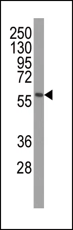
Western blot analysis of anti-PINK1 Monoclonal Antibody (PINK1 Mouse Monoclonal Antibody) in mouse brain tissue lysates. PINK1(arrow) was detected using the ascites Mab. (dilution 1:500)

Western blot analysis of anti-PINK1 Monoclonal Antibody (PINK1 Mouse Monoclonal Antibody) in mouse brain tissue lysates. PINK1(arrow) was detected using the ascites Mab. (dilution 1:500)

Catalog Number:MABN85907
Conjugate:Unconjugated
Size:100ul
Concentration:1mg/ml
Host:Mouse
Isotype:Mouse IgG1
Clone:Monoclonal
Immunogen:Recombinant PINK1 protein was used to produced this monoclonal antibody.
Reactivity:Human, Mouse
Applications:WB 1:500-1:2000,IHC 1:100-1:500
Molecular Weight:8kDa
Purification:Affinity purification
Form:liquid
Synonyms:Serine/threonine-protein kinase PINK1, mitochondrial, BRPK, PTEN-induced putative kinase protein 1, PINK1

This gene encodes a serine/threonine protein kinase that localizes to mitochondria. It is thought to protect cells from stress-induced mitochondrial dysfunction. Mutations in this gene cause one form of autosomal recessive early-onset Parkinson disease.

Applications

WB 1:500-1:2000,IHC 1:100-1:500

Immunogen

Recombinant PINK1 protein was used to produced this monoclonal antibody.
Target Background

This gene encodes a serine/threonine protein kinase that localizes to mitochondria. It is thought to protect cells from stress-induced mitochondrial dysfunction. Mutations in this gene cause one form of autosomal recessive early-onset Parkinson disease.

Synonyms:Serine/threonine-protein kinase PINK1, mitochondrial, BRPK, PTEN-induced putative kinase protein 1, PINK1

Storage

Store at 4°C short term. Aliquot and store at -20°C long term. Avoid freeze/thaw cycles.

Buffer

Purified antibody in PBS with 0.05% sodium azide.

Western blot analysis of anti-PINK1 Monoclonal Antibody (PINK1 Mouse Monoclonal Antibody) in mouse brain tissue lysates. PINK1(arrow) was detected using the ascites Mab. (dilution 1:500)

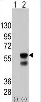
Western blot analysis of PINK (arrow) using mouse monoclonal PINK antibody(Ascites). 293 cell lysates (2 μg/lane) either nontransfected (Lane 1) or transiently transfected with the PINK gene (Lane 2) (Origene Technologies) (1:2000)

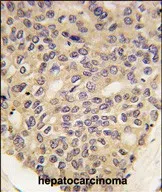
Formalin-fixed and paraffin-embedded human hepatocarcinoma tissue reacted with PINK1 Monoclonal Antibody (Cat.#AMM85907), which was peroxidase-conjugated to the secondary antibody, followed by DAB staining. This data demonstrates the use of this antibody for immunohistochemistry; clinical relevance has not been evaluated.

No documents available.