PLP1 Rabbit Polyclonal Antibody

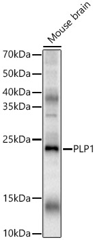
Western blot analysis of various lysates, using PLP1 Rabbit pAb  at 1:2000dilution.
Secondary antibody: HRP-conjugated Goat anti-Rabbit IgG (H+L) at 1:10000dilution.
Lysates/proteins: 25μg per lane.
Blocking buffer: 3% nonfat dry milk in TBST.
Detection: ECL West Pico Plus.
Exposure time: 30s.

Western blot analysis of various lysates, using PLP1 Rabbit pAb  at 1:2000dilution. Secondary antibody: HRP-conjugated Goat anti-Rabbit IgG (H+L) at 1:10000dilution. Lysates/proteins: 25μg per lane. Blocking buffer: 3% nonfat dry milk in TBST. Detection: ECL West Pico Plus. Exposure time: 30s.

Catalog Number:AB-83554
Conjugate:Unconjugated
Size:100 ug
Concentration:1mg/ml
Host:Rabbit
Isotype:IgG
Clone:POLY
Immunogen:Recombinant fusion protein containing a sequence corresponding to amino acids 90-150 of human PLP1.
Reactivity:Human,Mouse,Rat
Applications:Western Blot:   1:1000 - 1:5000 Immunohistochemistry (paraffin-embedded tissues):  1:50 - 1:200 Immunofluorescence:  1:50 - 1:200 Immunocytochemistry:   1:50 - 1:200 ELISA Recommended starting concentration is 1 μg/mL.
Molecular Weight:20-25kDa
Purification:Affinity purification
Form:Liquid
Synonyms:PLP; PMD; HLD1; MMPL; SPG2; GPM6C; PLP/DM20; PLP1

Applications

Western Blot:   1:1000 - 1:5000 Immunohistochemistry (paraffin-embedded tissues):  1:50 - 1:200 Immunofluorescence:  1:50 - 1:200 Immunocytochemistry:   1:50 - 1:200 ELISA Recommended starting concentration is 1 μg/mL.

Immunogen

Recombinant fusion protein containing a sequence corresponding to amino acids 90-150 of human PLP1.

Target Background

This gene encodes a transmembrane proteolipid protein that is the predominant component of myelin. The encoded protein may play a role in the compaction,stabilization, and maintenance of myelin sheaths, as well as in oligodendrocyte development and axonal survival. Mutations in this gene cause Pelizaeus-Merzbacher disease and spastic paraplegia type 2. Alternatively splicing results in multiple transcript variants, including the DM20 splice variant.

Synonyms:PLP; PMD; HLD1; MMPL; SPG2; GPM6C; PLP/DM20; PLP1

Storage

Store at -20℃. Avoid freeze / thaw cycles.

Buffer

PBS with 0.05% proclin300,50% glycerol,pH7.3.

Western blot analysis of various lysates, using PLP1 Rabbit pAb  at 1:2000dilution. Secondary antibody: HRP-conjugated Goat anti-Rabbit IgG (H+L) at 1:10000dilution. Lysates/proteins: 25μg per lane. Blocking buffer: 3% nonfat dry milk in TBST. Detection: ECL West Pico Plus. Exposure time: 30s.

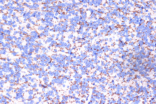
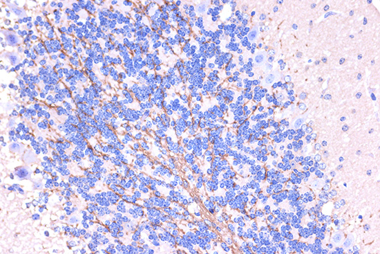
Immunohistochemistry analysis of paraffin-embedded Rat brain using PLP1 Rabbit pAb  at dilution of 1:100 (40x lens). Microwave antigen retrieval performed with 0.01M PBS Buffer (pH 7.2) prior to IHC staining.

Immunohistochemistry analysis of paraffin-embedded Mouse brain using PLP1 Rabbit pAb  at dilution of 1:100 (40x lens). Microwave antigen retrieval performed with 0.01M PBS Buffer (pH 7.2) prior to IHC staining.

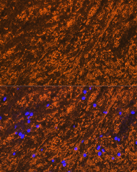
Immunofluorescence analysis of paraffin-embedded rat brain using PLP1 Rabbit pAb  at dilution of 1:100 (40x lens). Secondary antibody: Cy3-conjugated Goat anti- Rabbit IgG (H+L) at 1:500 dilution. Blue: DAPI for nuclear staining.

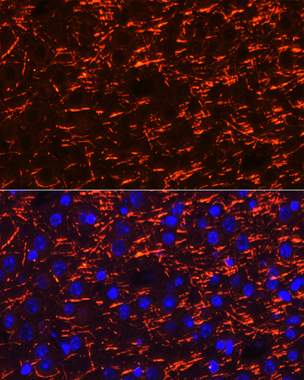
Immunofluorescence analysis of paraffin-embedded mouse brain using PLP1 Rabbit pAb  at dilution of 1:100 (40x lens). Secondary antibody: Cy3-conjugated Goat anti-Rabbit IgG (H+L) at 1:500 dilution. Blue: DAPI for nuclear staining.

No documents available.