PPAR-γ Rabbit Polyclonal Antibody

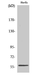
Western Blot analysis of HuvEc cells using PPAR-γ Polyclonal Antibody diluted at 1:1000

Western Blot analysis of HuvEc cells using PPAR-γ Polyclonal Antibody diluted at 1:1000

Catalog Number:AB-E12636
Conjugate:Unconjugated
Size:100 ug
Concentration:1mg/ml
Host:Rabbit
Isotype:IgG
Clone:POLY
Immunogen:The antiserum was produced against synthesized peptide derived from human PPAR-gamma. AA range:78-127.
Reactivity:Human;Mouse;Rat
Applications:<p style="text-align: left;">Western Blot:  1:500-1:2000                                                              Immunofluorescence:   1:50-200 Immunohistochemistry: 1/100 - 1/300.</p>
Molecular Weight:57kD
Purification:The antibody was affinity-purified from rabbit antiserum by affinity-chromatography using epitope-specific immunogen.
Form:Liquid
Synonyms:PPARG; NR1C3; Peroxisome proliferator-activated receptor gamma; PPAR-gamma; Nuclear receptor subfamily 1 group C member 3

Applications

<p style="text-align: left;">Western Blot:  1:500-1:2000                                                              Immunofluorescence:   1:50-200 Immunohistochemistry: 1/100 - 1/300.</p>

Immunogen

The antiserum was produced against synthesized peptide derived from human PPAR-gamma. AA range:78-127.

Target Background

Peroxisome proliferator activated receptor gamma(PPARG) Homo sapiens This gene encodes a member of the peroxisome proliferator-activated receptor (PPAR) subfamily of nuclear receptors. PPARs form heterodimers with retinoid X receptors (RXRs) and these heterodimers regulate transcription of various genes. Three subtypes of PPARs are known: PPAR-alpha, PPAR-delta, and PPAR-gamma. The protein encodedby this gene is PPAR-gamma and is a regulator of adipocyte differentiation. Additionally, PPAR-gamma has been implicated in the pathology of numerous diseases including obesity, diabetes, atherosclerosis and cancer. Alternatively spliced transcript variants that encode different isoforms have been described.

Synonyms:PPARG; NR1C3; Peroxisome proliferator-activated receptor gamma; PPAR-gamma; Nuclear receptor subfamily 1 group C member 3

Storage

Store at -20°C. Avoid repeated freeze-thaw cycles.

Buffer

Liquid in PBS containing 50% glycerol, 0.5% BSA and 0.02% sodium azide.

Western Blot analysis of HuvEc cells using PPAR-γ Polyclonal Antibody diluted at 1:1000

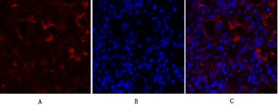
Immunofluorescence analysis of rat-lung tissue. 1,PPAR-γ Polyclonal Antibody(red) was diluted at 1:200(4°C,overnight). 2, Cy3 labled Secondary antibody was diluted at 1:300(room temperature, 50min).3, Picture B: DAPI(blue) 10min. Picture A:Target. Picture

Immunofluorescence analysis of rat-lung tissue. 1,PPAR-γ Polyclonal Antibody(red) was diluted at 1:200(4°C,overnight). 2, Cy3 labled Secondary antibody was diluted at 1:300(room temperature, 50min).3, Picture B: DAPI(blue) 10min. Picture A:Target. Picture B: DAPI. Picture C: merge of A+B

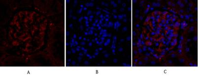
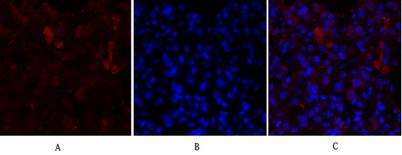
Immunofluorescence analysis of rat-kidney tissue. 1,PPAR-γ Polyclonal Antibody(red) was diluted at 1:200(4°C,overnight). 2, Cy3 labled Secondary antibody was diluted at 1:300(room temperature, 50min).3, Picture B: DAPI(blue) 10min. Picture A:Target. Picture B: DAPI. Picture C: merge of A+B

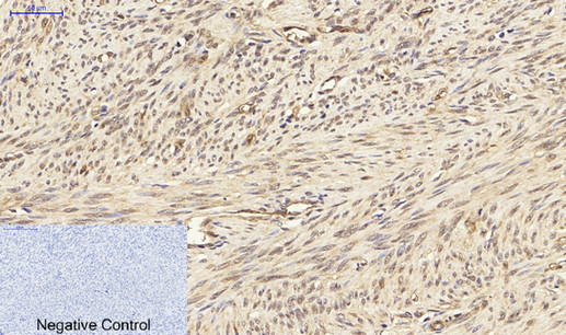
Immunohistochemical analysis of paraffin-embedded Human-uterus tissue. 1,PPAR-γ Polyclonal Antibody was diluted at 1:200(4°C,overnight). 2, Sodium citrate pH 6.0 was used for antibody retrieval(&gt;98°C,20min). 3,Secondary antibody was diluted at 1:200(room tempeRature, 30min). Negative control was used by secondary antibody only.

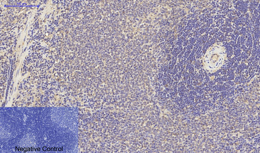
Immunohistochemical analysis of paraffin-embedded Rat-spleen tissue. 1,PPAR-γ Polyclonal Antibody was diluted at 1:200(4°C,overnight). 2, Sodium citrate pH 6.0 was used for antibody retrieval(&gt;98°C,20min). 3,Secondary antibody was diluted at 1:200(room tempeRature, 30min). Negative control was used by secondary antibody only.

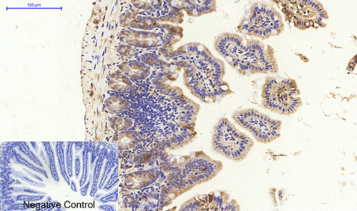
Immunohistochemical analysis of paraffin-embedded Mouse-colon tissue. 1,PPAR-γ Polyclonal Antibody was diluted at 1:200(4°C,overnight). 2, Sodium citrate pH 6.0 was used for antibody retrieval(&gt;98°C,20min). 3,Secondary antibody was diluted at 1:200(room tempeRature, 30min). Negative control was used by secondary antibody only.

No documents available.