RUNX2

Western Blot on 3T3 cells

Western Blot on 3T3 cells

Catalog Number:AB-84116
Size:100 ul
Concentration:1mg/ml
Host:Rb
Isotype:IgG
Clone:POLY
Immunogen:Synthesized peptide derived from human RUNX2.
Reactivity:Hu, Ms
Applications:Western Blot: 1:500 - 1:2000 Immunofluorescence: 1:50-1:200. ELISA: 1/2000. Not yet tested in other applications
Molecular Weight:56kDa
Purification:Aff. Pur.
Form:Liquid

Applications

Western Blot: 1:500 - 1:2000 Immunofluorescence: 1:50-1:200. ELISA: 1/2000. Not yet tested in other applications

Immunogen

Synthesized peptide derived from human RUNX2.
Target Background

Transcription factor involved in osteoblastic differentiation and skeletal morphogenesis. Essential for the maturation of osteoblasts and both intramembranous and endochondral ossification. CBF binds to the core site, 5'-PYGPYGGT-3', of a number of enhancers and promoters, including murine leukemia virus, polyomavirus enhancer, T-cell receptor enhancers, osteocalcin, osteopontin, bone sialoprotein, alpha 1(I) collagen, LCK, IL-3 and GM-CSF promoters. In osteoblasts, supports transcription activation: synergizes with SPEN/MINT to enhance FGFR2-mediated activation of the osteocalcin FGF- responsive element (OCFRE) . Inhibits KAT6B-dependent transcriptional activation. Modifications: Phosphorylated; probably by MAP kinases (MAPK). Phosphorylation by HIPK3 is required for the SPEN/MINT and FGF2 transactivation during osteoblastic differentiation . Phosphorylation at Ser-451 by CDK1 promotes endothelial cell proliferation required for tumor angiogenesis probably by facilitating cell cycle progression. Isoform 3 is phosphorylated on Ser-340.

Storage

At +4°C for short term. For longer term store at -20°C, avoid repeat freeze-thaw cycles.

Buffer

Liquid in PBS containing 50% glycerol, 0.5% BSA and 0.02% sodium azide.

Western Blot on 3T3 cells

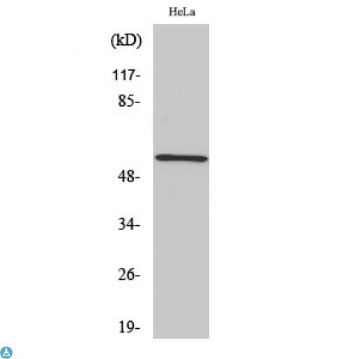
Western blot on Hela cells

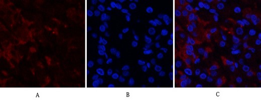
Immunofluorescence analysis of human-stomach tissue. 1,RUNX2 Polyclonal Antibody(red) was diluted at 1:200(4°C,overnight). 2, Cy3 labled Secondary antibody was diluted at 1:300(room temperature, 50min).3, Picture B: DAPI(blue) 10min. Picture A:Target. Picture B: DAPI. Picture C: merge of A+B

No documents available.