SDHB

Western blot analysis of extracts of 
various cell lines, using SDHB  antibody
 at 1:1000 dilution.
Secondary antibody: HRP 
Goat Anti-Rabbit IgG (H+L) at 
1:10000 dilution.
Lysates/proteins: 25ug per lane.
Blocking buffer: 3% nonfat dry milk in TBST.
Detection: ECL West Pico Plus.Exposure time: 1s.

Western blot analysis of extracts of various cell lines, using SDHB antibody at 1:1000 dilution. Secondary antibody: HRP Goat Anti-Rabbit IgG (H+L) at 1:10000 dilution. Lysates/proteins: 25ug per lane. Blocking buffer: 3% nonfat dry milk in TBST. Detection: ECL West Pico Plus.Exposure time: 1s.

Catalog Number:MAB-94613
Size:100 ug
Concentration:1mg/ml
Host:Rb
Isotype:IgG
Clone:21A11AE7
Immunogen:Recombinant fusion protein containing a sequence corresponding to amino acids 29-280 of human SDHB.
Reactivity:Hu, Ms, Rt
Applications:Western Blot: 1:500 - 1:2000 Immunohistochemistry: 1:50-1:100 Immunofluorescence: 1:50-1:200
Molecular Weight:32kDa
Purification:Aff. Pur
Form:Liquid
Synonyms:CWS2; IP; PGL4; SDH; SDH1; SDH2; SDHIP

Applications

Western Blot: 1:500 - 1:2000 Immunohistochemistry: 1:50-1:100 Immunofluorescence: 1:50-1:200

Immunogen

Recombinant fusion protein containing a sequence corresponding to amino acids 29-280 of human SDHB.

Target Background

Complex II of the respiratory chain, which is specifically involved in the oxidation of succinate, carries electrons from FADH to CoQ. The complex is composed of four nuclear-encoded subunits and is localized in the mitochondrial inner membrane. The iron-sulfur subunit is highly conserved and contains three cysteine-rich clusters which may comprise the iron-sulfur centers of the enzyme. Sporadic and familial mutations in this gene result in paragangliomas and pheochromocytoma, and support a link between mitochondrial dysfunction and tumorigenesis.

Synonyms:CWS2; IP; PGL4; SDH; SDH1; SDH2; SDHIP

Storage

Store at -20℃. Avoid freeze / thaw cycles.

Buffer

PBS with 0.02% sodium azide,50% glycerol,pH7.3.

Western blot analysis of extracts of various cell lines, using SDHB antibody at 1:1000 dilution. Secondary antibody: HRP Goat Anti-Rabbit IgG (H+L) at 1:10000 dilution. Lysates/proteins: 25ug per lane. Blocking buffer: 3% nonfat dry milk in TBST. Detection: ECL West Pico Plus.Exposure time: 1s.

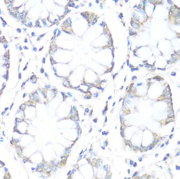
Immunohistochemistry of paraffin embedded human colon using SDHB antibody at dilution of 1:100 (40x lens).

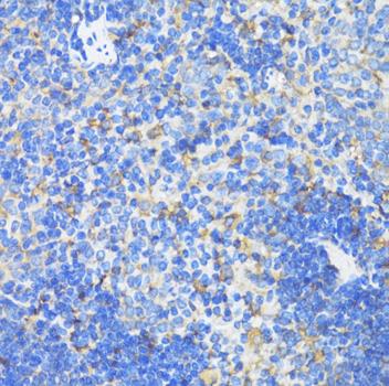
Immunohistochemistry of paraffin embedded mouse spleen using SDHB antibody at dilution of 1:100 (40x lens).

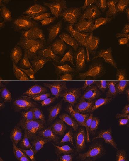
Immunofluorescence analysis of U2OS cells using SDHB Rabbit mAb at dilution of 1:100. Blue: DAPI for nuclear staining.

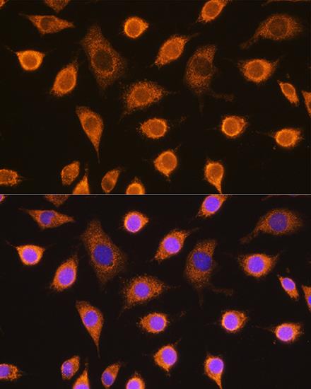
Immunofluorescence analysis of L929 cells using SDHB Rabbit mAb at dilution of 1:100. Blue: DAPI for nuclear staining.

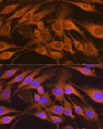
Immunofluorescence analysis of C6 cells using SDHB Rabbit mAb at dilution of 1:100. Blue: DAPI for nuclear staining.

No documents available.