
SOD1

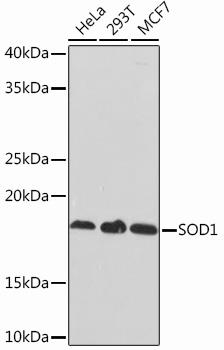
Western blot analysis of extracts of various cell lines, using SOD1 Rabbit mAb at 1:1000 dilution.
Secondary antibody: HRP Goat Anti-Rabbit IgG
(H+L) at 1:10000 dilution.
Lysates/proteins: 25ug per lane.
Blocking buffer: 3% nonfat dry milk in TBST.
Detection: ECL West Pico Plus. 
Exposure time: 1s.

Western blot analysis of extracts of various cell lines, using SOD1 Rabbit mAb at 1:1000 dilution. Secondary antibody: HRP Goat Anti-Rabbit IgG (H+L) at 1:10000 dilution. Lysates/proteins: 25ug per lane. Blocking buffer: 3% nonfat dry milk in TBST. Detection: ECL West Pico Plus. Exposure time: 1s.

Catalog Number:MAB-94600
Size:100 ug
Concentration:1mg/ml
Host:Rb
Isotype:IgG
Clone:SO-F
Immunogen:A synthesized peptide derived from human SOD1
Reactivity:Hu, Rt, Ms
Applications:Western Blot: 1:500 - 1:2000
Molecular Weight:16kDa
Purification:Aff. Pur.
Form:Liquid
Synonyms:ALS; ALS1; HEL-S-44; IPOA; SOD; hSod1; homodimer

Applications

Western Blot: 1:500 - 1:2000

Immunogen

A synthesized peptide derived from human SOD1
Target Background

The protein encoded by this gene binds copper and zinc ions and is one of two isozymes responsible for destroying free superoxide radicals in the body. The encoded isozyme is a soluble cytoplasmic protein, acting as a homodimer to convert naturally-occuring but harmful superoxide radicals to molecular oxygen and hydrogen peroxide. The other isozyme is a mitochondrial protein. Mutations in this gene have been implicated as causes of familial amyotrophic lateral sclerosis. Rare transcript variants have been reported for this gene.

Synonyms:ALS; ALS1; HEL-S-44; IPOA; SOD; hSod1; homodimer

Storage

Store at -20℃. Avoid freeze / thaw cycles.

Buffer

PBS with 0.02% sodium azide,0.05% BSA,50% glycerol,pH7.3.

Western blot analysis of extracts of various cell lines, using SOD1 Rabbit mAb at 1:1000 dilution. Secondary antibody: HRP Goat Anti-Rabbit IgG (H+L) at 1:10000 dilution. Lysates/proteins: 25ug per lane. Blocking buffer: 3% nonfat dry milk in TBST. Detection: ECL West Pico Plus. Exposure time: 1s.

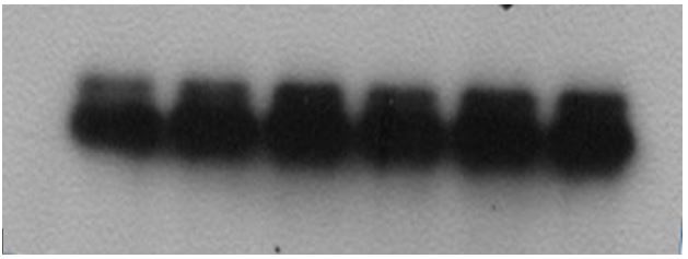
Western blot analysis of extracts of various cell lines, using SOD1 Rabbit mAb at 1:1000 dilution. Secondary antibody: HRP Goat Anti-Rabbit IgG (H+L) at 1:10000 dilution. Lysates/proteins: 25ug per lane. Blocking buffer: 3% nonfat dry milk in TBST. Detection: ECL West Pico Plus. Exposure time: 10s.

Review for SOD1 Rabbit mAb Experiment Type Western blot(WB)

No documents available.