
SQSTM1 / P62

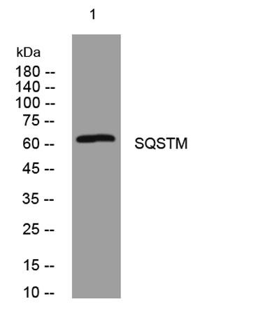
Anti-SQSTM1/p62 antibody, Western blotting
Lane 1: Rat Brain Tissue Lysate
Lane 2: HELA Cell Lysate
Lane 3: U87 Cell Lysate
Lane 4: A549 Cell Lysate

Anti-SQSTM1/p62 antibody, Western blotting Lane 1: Rat Brain Tissue Lysate Lane 2: HELA Cell Lysate Lane 3: U87 Cell Lysate Lane 4: A549 Cell Lysate

Catalog Number:AB-J14285
Size:100 ug
Concentration:1mg/ml
Host:Rb
Isotype:IgG
Clone:POLY
Immunogen:Synthesized peptide derived from human SQSTM
Reactivity:Hu, Ms, Rt
Applications:Western Blot: 1:500-2000 Immunofluorescence: 1:100-500 Immunocytochemistry: 1:100-500
Molecular Weight:62kDa
Purification:Aff. Pur.
Form:Liquid

Applications

Western Blot: 1:500-2000 Immunofluorescence: 1:100-500 Immunocytochemistry: 1:100-500

Immunogen

Synthesized peptide derived from human SQSTM
Target Background

SQSTM1(Sequestosome-1), also known as Ubiquitin-Binding Protein P62 or P62, is a protein that in humans is encoded by the SQSTM1 gene. The Src homology type 2(SH2) domain is a highly conserved motif of about 100 amino acids which mediates protein-protein interactions by binding to phosphotyrosine.p56-lck, a T-cell-specific src family tyrosine kinase with an SH2 domain, is involved in T-cell signal transduction. The International Radiation Hybrid Mapping Consortium mapped the p62 gene to chromosome 5q35. Park et al.(1995) found that the p56-lck SH2 domain binds to p62 at the ser59 of p62 only when that serine is phosphorylated. Joung et al.(1996) expressed epitope-tagged p62 in Hela cells and showed that the expressed protein bound to the lck SH2 domain and that this binding was dependent on the N-terminal 50 amino acids of p62 but not on the tyrosine residue in this region.

Storage

At -20˚C for one year. at 4˚C for one month. It can also be aliquotted and stored frozen at -20˚C for a longer time.Avoid repeated freezing and thawing.

Buffer

Liquid in PBS containing 50% glycerol, 0.5% BSA and 0.02% sodium azide.

Anti-SQSTM1/p62 antibody, Western blotting Lane 1: Rat Brain Tissue Lysate Lane 2: HELA Cell Lysate Lane 3: U87 Cell Lysate Lane 4: A549 Cell Lysate

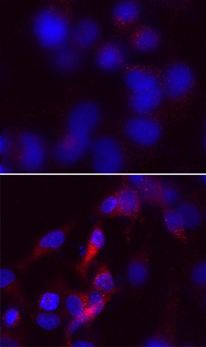
Immunofluorescence analysis of Hela cells treated or un-treated with UV 30MIN. 1,P62 Polyclonal Antibody(red) was diluted at 1:200(4°C,overnight). 2, AFlour 594 labled Secondary antibody was diluted at 1:300(room temperature, 50min).

No documents available.