TBX21 /T-bet

Figure 1. Western blot analysis of TBX21 using anti-TBX21
Antibody.    
Electrophoresis was performed on a 5-20% SDS-PAGE gel
at 70V (Stacking gel) / 90V (Resolving gel) for 2-3 hours. The
sample well of each lane was loaded with 50ug of sample
under reducing conditions.
Lane 1: mouse RAW246.7 whole cell lysates.
After Electrophoresis, proteins were transferred to a
Nitrocellulose membrane at 150mA for 50-90 minutes.
Blocked the membrane with 5% Non-fat Milk/ TBS for 1.5
hour at RT. The membrane was incubated with rabbit anti-
TBX21 antigen affinity purified polyclonal antibody at 0.5 mug/mL overnight at 4°C, then washed
with TBS-0.1%Tween 3 times with 5 minutes each and
probed with a goat anti-rabbit IgG-HRP secondary antibody
at a dilution of 1:5000 for 1.5 hour at RT. The signal is
developed using an Enhanced Chemiluminescent detection
(ECL) kit  with Tanon 5200 system. 
A specific band was detected for TBX21 at approximately
65KD. The expected band size for TBX21 is at 58KD.

Figure 1. Western blot analysis of TBX21 using anti-TBX21 Antibody. Electrophoresis was performed on a 5-20% SDS-PAGE gel at 70V (Stacking gel) / 90V (Resolving gel) for 2-3 hours. The sample well of each lane was loaded with 50ug of sample under reducing conditions. Lane 1: mouse RAW246.7 whole cell lysates. After Electrophoresis, proteins were transferred to a Nitrocellulose membrane at 150mA for 50-90 minutes. Blocked the membrane with 5% Non-fat Milk/ TBS for 1.5 hour at RT. The membrane was incubated with rabbit anti- TBX21 antigen affinity purified polyclonal antibody at 0.5 mug/mL overnight at 4°C, then washed with TBS-0.1%Tween 3 times with 5 minutes each and probed with a goat anti-rabbit IgG-HRP secondary antibody at a dilution of 1:5000 for 1.5 hour at RT. The signal is developed using an Enhanced Chemiluminescent detection (ECL) kit with Tanon 5200 system. A specific band was detected for TBX21 at approximately 65KD. The expected band size for TBX21 is at 58KD.

Catalog Number:AB-84321
Size:100ug
Concentration:1mg/ml
Host:Rb
Isotype:IgG
Clone:POLY
Immunogen:A synthetic peptide corresponding to a sequence of human Tbet/ Tbx21(NNVTQMIVLQSLHKYQPRLHIVEVNDGE).
Reactivity:Hu, Ms, Rt
Applications:Western blot: 0.25-0.5ug/ml Flow Cytometry: 1-3ug/1x106 cells,
Molecular Weight:58kDa
Purification:Aff. Pur.
Form:Liquid

Applications

Western blot: 0.25-0.5ug/ml Flow Cytometry: 1-3ug/1x106 cells,

Immunogen

A synthetic peptide corresponding to a sequence of human Tbet/ Tbx21(NNVTQMIVLQSLHKYQPRLHIVEVNDGE).

Target Background

T-box transcription factor TBX21 is a protein that in humans is encoded by the TBX21 gene. It is mapped to 17q21.32. This gene is a member of a phylogenetically conserved family of genes that share a common DNA-binding domain, the T-box. T-box genes encode transcription factors involved in the regulation of developmental processes. This gene is the human ortholog of mouse Tbx21/Tbet gene. Studies in mouse show that Tbx21 protein is a Th1 cell-specific transcription factor that controls the expression of the hallmark Th1 cytokine, interferon-gamma (IFNG). Expression of the human ortholog also correlates with IFNG expression in Th1 and natural killer cells, suggesting a role for this gene in initiating Th1 lineage development from naive Th precursor cells.

Storage

At -20°C for one year. After reconstitution, at 4°C for one month. It can also be aliquotted and stored frozen at -20°C for a longer time. Avoid repeated freezing and thawing.

Buffer

Each vial contains 4mg Trehalose, 0.9mg NaCl, 0.2mg Na2HPO4, 0.05mg NaN3.

Figure 1. Western blot analysis of TBX21 using anti-TBX21 Antibody. Electrophoresis was performed on a 5-20% SDS-PAGE gel at 70V (Stacking gel) / 90V (Resolving gel) for 2-3 hours. The sample well of each lane was loaded with 50ug of sample under reducing conditions. Lane 1: mouse RAW246.7 whole cell lysates. After Electrophoresis, proteins were transferred to a Nitrocellulose membrane at 150mA for 50-90 minutes. Blocked the membrane with 5% Non-fat Milk/ TBS for 1.5 hour at RT. The membrane was incubated with rabbit anti- TBX21 antigen affinity purified polyclonal antibody at 0.5 mug/mL overnight at 4°C, then washed with TBS-0.1%Tween 3 times with 5 minutes each and probed with a goat anti-rabbit IgG-HRP secondary antibody at a dilution of 1:5000 for 1.5 hour at RT. The signal is developed using an Enhanced Chemiluminescent detection (ECL) kit with Tanon 5200 system. A specific band was detected for TBX21 at approximately 65KD. The expected band size for TBX21 is at 58KD.

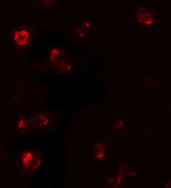
Immunofluorescent analysis of 293 cells labeling T-bet / Tbx21 with AB-84247 at 5 µg/ml.

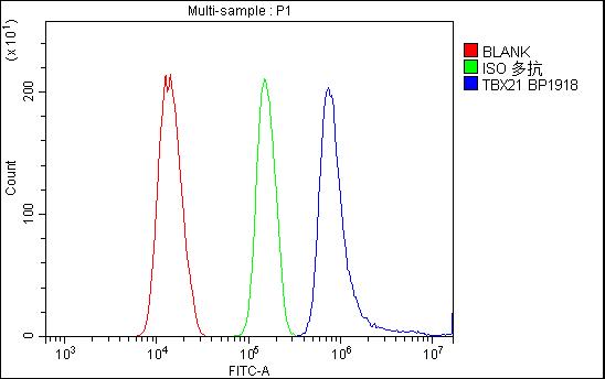
Figure 2. Flow Cytometry analysis of HEPA1-6 cells using anti- TBX21 antibody. Overlay histogram showing HEPA1-6 cells stained with (Blue line).The cells were blocked with 10% normal goat serum. And then incubated with rabbit anti-TBX21 Antibody 1mug/1x106 cells) for 30 min at 20°C. DyLight®488 conjugated goat anti-rabbit IgG (5-10mug/1x106 cells) was used as secondary antibody for 30 minutes at 20°C. Isotype control antibody (Green line) was rabbit IgG (1mug/1x106) used under the same conditions. Unlabelled sample (Red line) was also used as a control.

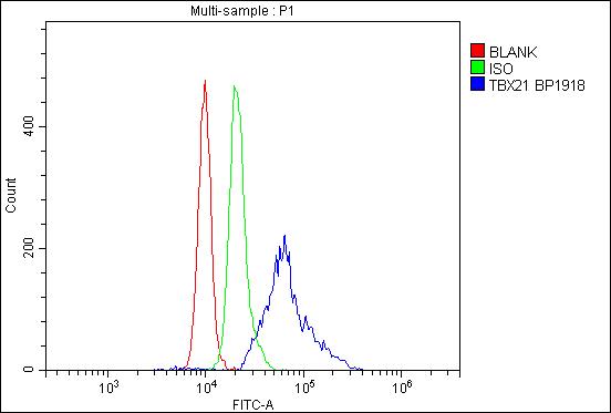
Figure 3. Flow Cytometry analysis of NRK cells using anti- TBX21 antibody. Overlay histogram showing NRK cells stained with (Blue line).The cells were blocked with 10% normal goat serum. And then incubated with rabbit anti-TBX21 Antibody (1mug/1x106 cells) for 30 min at 20°C. DyLight®488 conjugated goat anti-rabbit IgG (5-10mug/1x106 cells) was used as secondary antibody for 30 minutes at 20°C. Isotype control antibody (Green line) was rabbit IgG (1mug/1x106) used under the same conditions. Unlabelled sample (Red line) was also used as a control

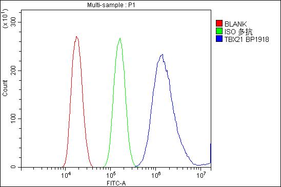
Figure 4. Flow Cytometry analysis of mouse PBMC cells using anti-TBX21 antibody Overlay histogram showing mouse PBMC cells stained with (Blue line).The cells were blocked with 10% normal goat serum. And then incubated with rabbit anti-TBX21 Antibody (1mug/1x106 cells) for 30 min at 20°C. DyLight®488 conjugated goat anti-rabbit IgG (5-10mug/1x106 cells) was used as secondary antibody for 30 minutes at 20°C. Isotype control antibody (Green line) was rabbit IgG (1mug/1x106) used under the same conditions. Unlabelled sample (Red line) was also used as a control.

No documents available.