TGF beta1

Western blot analysis of extracts of
various cell lines, using TGFBI
antibody  at 1:1000 dilution.
Secondary antibody: HRP Goat Anti-
Rabbit IgG (H+L) at 1:10000
dilution.
Lysates/proteins: 25ug per lane.
Blocking buffer: 3% nonfat dry milk in
TBST.
Detection: ECL Basic Kit. 
Exposure time: 5s.

Western blot analysis of extracts of various cell lines, using TGFBI antibody at 1:1000 dilution. Secondary antibody: HRP Goat Anti- Rabbit IgG (H+L) at 1:10000 dilution. Lysates/proteins: 25ug per lane. Blocking buffer: 3% nonfat dry milk in TBST. Detection: ECL Basic Kit. Exposure time: 5s.

Catalog Number:AB-84169
Size:100 ug
Concentration:1mg/ml
Host:Rb
Isotype:IgG
Clone:POLY
Immunogen:Recombinant fusion protein containing a sequence corresponding to amino acids 481-683 of human TGFBI
Reactivity:Hu,Ms
Applications:Western Blot: 1:500 - 1:2000 Immunohistochemistry: 1:50 - 1:200
Molecular Weight:75kDa
Purification:Aff. Pur.
Form:Liquid
Synonyms:TGFBI; BIGH3; CDB1; CDG2; CDGG1; CSD; CSD1; CS D2; CSD3; EBMD; LCD1; transforming growth fact or beta induced.

Applications

Western Blot: 1:500 - 1:2000 Immunohistochemistry: 1:50 - 1:200

Immunogen

Recombinant fusion protein containing a sequence corresponding to amino acids 481-683 of human TGFBI

Target Background

This gene encodes an RGD-containing protein that binds to type I, II and IV collagens. The RGD motif is found in many extracellular matrix proteins modulating cell adhesion and serves as a ligand recognition sequence for several integrins. This protein plays a role in cell-collagen interactions and may be involved in endochondrial bone formation in cartilage. The protein is induced by transforming growth factor-beta and acts to inhibit cell adhesion. Mutations in this gene are associated with multiple types of corneal dystrophy.

Synonyms:TGFBI; BIGH3; CDB1; CDG2; CDGG1; CSD; CSD1; CS D2; CSD3; EBMD; LCD1; transforming growth fact or beta induced.

Storage

Store at -20℃. Avoid freeze / thaw cycles.

Buffer

PBS with 0.02% sodium azide,50% glycerol,pH7.3.

Western blot analysis of extracts of various cell lines, using TGFBI antibody at 1:1000 dilution. Secondary antibody: HRP Goat Anti- Rabbit IgG (H+L) at 1:10000 dilution. Lysates/proteins: 25ug per lane. Blocking buffer: 3% nonfat dry milk in TBST. Detection: ECL Basic Kit. Exposure time: 5s.

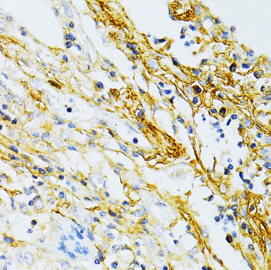
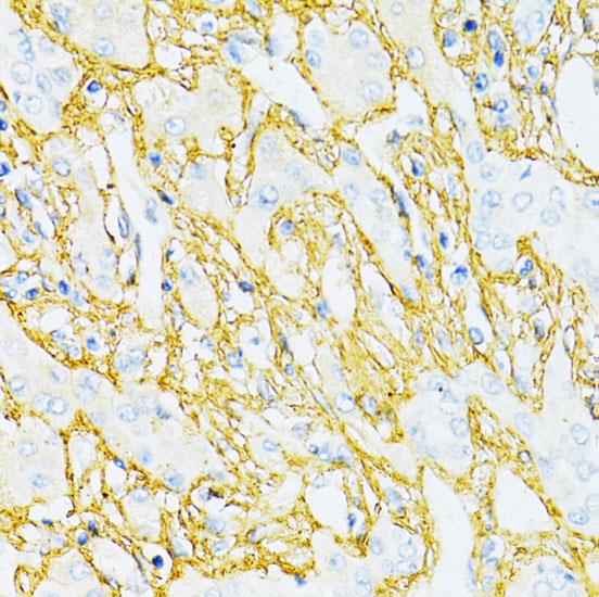
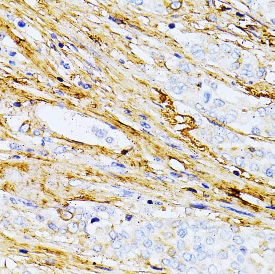
Immunohistochemistry of paraffin embedded human liver cancer using TGFBI antibody at dilution of 1:100 (40x lens).

Immunohistochemistry of paraffin embedded human colon carcinoma using TGFBI antibody at dilution of 1:100 (40x lens).

Immunohistochemistry of paraffin embedded human lung cancer using TGFBI antibody at dilution of 1:100 (40x lens).

No documents available.