
TGS101

Western blot analysis of TSG101 expression in Jurkat cell lysate 
Electrophoresis was performed on a 5-20% SDS-PAGE gel at 70V (Stacking gel) / 90V (Resolving gel) for 2-3 hours. 
50ug of sample  was loaded each laneunder reducing conditions.
After Electrophoresis, proteins were transferred to a NC  at 150mA for 50-90 minutes. Blocked the membrane with 5% Non-fat Milk/ TBS for 1.5 hour at RT. The membrane was incubated   overnight at 4°C, then washed with TBS-0.1%Tween 3 times with 5 minutes each and probed with a goat anti-rabbit IgG-HRP secondary antibody at a dilution of 1:10.000 for 1.5 hour at RT.                                                                                 ECL West Pico Plus with Tanon 5200 system.

Western blot analysis of TSG101 expression in Jurkat cell lysate Electrophoresis was performed on a 5-20% SDS-PAGE gel at 70V (Stacking gel) / 90V (Resolving gel) for 2-3 hours. 50ug of sample was loaded each laneunder reducing conditions. After Electrophoresis, proteins were transferred to a NC at 150mA for 50-90 minutes. Blocked the membrane with 5% Non-fat Milk/ TBS for 1.5 hour at RT. The membrane was incubated overnight at 4°C, then washed with TBS-0.1%Tween 3 times with 5 minutes each and probed with a goat anti-rabbit IgG-HRP secondary antibody at a dilution of 1:10.000 for 1.5 hour at RT. ECL West Pico Plus with Tanon 5200 system.

Catalog Number:MAB-94384
Conjugate:Unconjugated
Size:100 ug
Concentration:1mg/ml
Host:Rb
Isotype:IgG
Clone:IDG-20
Immunogen:A synthesized peptide derived from human TSG101
Reactivity:Hu, Ms, Rt
Applications:Western Blot: 1:500-1:2000 Immunohistochemistry: 1:50-1:200 ICC/IF 1:50-1:200 Flow Cytometry 1:50
Molecular Weight:43kDa
Purification:Aff. Pur.
Form:Liquid
Synonyms:Tumor susceptibility gene 101 protein;ESCRT-I complex subunit TSG101;TSG101;

Applications

Western Blot: 1:500-1:2000 Immunohistochemistry: 1:50-1:200 ICC/IF 1:50-1:200 Flow Cytometry 1:50

Immunogen

A synthesized peptide derived from human TSG101
Target Background

Component of the ESCRT-I complex, a regulator of vesicular trafficking process. Binds to ubiquitinated cargo proteins and is required for the sorting of endocytic ubiquitinated cargos into multivesicular bodies (MVBs). Mediates the association between the ESCRT-0 and ESCRT-I complex. Required for completion of cytokinesis; the function requires CEP55. May be involved in cell growth and differentiation. Acts as a negative growth regulator. Involved in the budding of many viruses through an interaction with viral proteins that contain a late-budding motif P-[ST]-A-P. This interaction is essential for viral particle budding of numerous retroviruses..Subcellular Localization: Cytoplasm. Membrane; Peripheral membrane protein. Nucleus. Late endosome membrane; Peripheral membrane protein. Mainly cytoplasmic.Membrane-associated when active and soluble when inactive. Depending on the stage of the cell cycle, detected in the nucleus. Colocalized with CEP55 in the midbody during cytokinesis.

Synonyms:Tumor susceptibility gene 101 protein;ESCRT-I complex subunit TSG101;TSG101;

Storage

Store for short term at 4-8°C .At -20°C for longer term. Avoid freeze / thaw cycles.

Buffer

PBS with 0.02% sodium azide, 50% glycerol, pH7.3.

Western blot analysis of TSG101 expression in Jurkat cell lysate Electrophoresis was performed on a 5-20% SDS-PAGE gel at 70V (Stacking gel) / 90V (Resolving gel) for 2-3 hours. 50ug of sample was loaded each laneunder reducing conditions. After Electrophoresis, proteins were transferred to a NC at 150mA for 50-90 minutes. Blocked the membrane with 5% Non-fat Milk/ TBS for 1.5 hour at RT. The membrane was incubated overnight at 4°C, then washed with TBS-0.1%Tween 3 times with 5 minutes each and probed with a goat anti-rabbit IgG-HRP secondary antibody at a dilution of 1:10.000 for 1.5 hour at RT. ECL West Pico Plus with Tanon 5200 system.

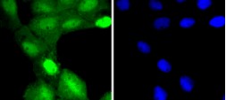
IF analysis of immunocytochemical section of A431 cell using anti- TSG101 antibody Enzyme antigen retrieval was performed using IHC enzyme antigen retrieval reagent for 15 mins. The tissue section was blocked with 10% goat serum. The tissue section was then incubated with 2μg/mL rabbit anti- TSG101 Antibody overnight at 4°C. AF488 Conjugated Goat Anti-Rabbit IgG was used as secondary antibody at 1:100 dilution and incubated for 30 minutes at 37°C. The section was counterstained with DAPI. Visualize using a fluorescence microscope and filter sets appropriate for the label use

No documents available.