TIMP-1 Rabbit Polyclonal Antibody

Western blot analysis of lysates from 1) SH-SY5Y, 2) 293 , 3) rat brain cells, (Green) primary antibody was diluted
at 1:1000, 4°over night, secondary antibody was diluted at 1:10000, 37° 1hour. 
(Red) Actin β Monoclonal Antibody(5B7)  antibody was diluted at 1:5000 as loading control, 4° over night,secondary antibodywas diluted at 1:10000, 37° 1hour.

Western blot analysis of lysates from 1) SH-SY5Y, 2) 293 , 3) rat brain cells, (Green) primary antibody was diluted at 1:1000, 4°over night, secondary antibody was diluted at 1:10000, 37° 1hour. (Red) Actin β Monoclonal Antibody(5B7) antibody was diluted at 1:5000 as loading control, 4° over night,secondary antibodywas diluted at 1:10000, 37° 1hour.

Catalog Number:AB-10132
Conjugate:Unconjugated
Size:100 ug
Concentration:1mg/ml
Host:Rabbit
Isotype:IgG
Clone:POLY
Immunogen:The antiserum was produced against synthesized peptide derived from human TIMP1. AA range:61-110
Reactivity:Human;Mouse;Rat, Rabbit
Applications:Western Blot: 1/500 - 1/2000 Immunofluorescence: 1:50-200 ELISA: 1/10000
Molecular Weight:24kD
Purification:The antibody was affinity-purified from rabbit antiserum by affinity-chromatography using epitope-specific immunogen.
Form:Liquid

Applications

Western Blot: 1/500 - 1/2000 Immunofluorescence: 1:50-200 ELISA: 1/10000

Immunogen

The antiserum was produced against synthesized peptide derived from human TIMP1. AA range:61-110
Target Background

This gene belongs to the TIMP gene family. The proteins encoded by this gene family are natural inhibitors of the matrix metalloproteinases (MMPs), a group of peptidases involved in degradation of theextracellular matrix. In addition to its inhibitory role against most of the known MMPs, the encoded protein is able to promote cell proliferation in a wide range of cell types, and may also have an anti-apoptotic function. Transcription of this gene is highly inducible in response to many cytokines and hormones. In addition, the expression from some but not all inactive X chromosomes suggests that this gene inactivation is polymorphic in human females. This gene is located within intron 6 of the synapsin I gene and is transcribed in the opposite direction.

Storage

Store at -20°C. Avoid repeated freeze-thaw cycles.

Buffer

Liquid in PBS containing 50% glycerol, 0.5% BSA and 0.02% sodium azide.

Western blot analysis of lysates from 1) SH-SY5Y, 2) 293 , 3) rat brain cells, (Green) primary antibody was diluted at 1:1000, 4°over night, secondary antibody was diluted at 1:10000, 37° 1hour. (Red) Actin β Monoclonal Antibody(5B7) antibody was diluted at 1:5000 as loading control, 4° over night,secondary antibodywas diluted at 1:10000, 37° 1hour.

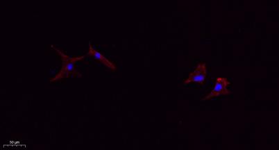
Immunofluorescence analysis of A549. 1,primary Antibody(red) was diluted at 1:200(4°C overnight). 2, Goat Anti Rabbit IgG (H&L) - Alexa Fluor 594 Secondary antibody was diluted at 1:1000(room temperature, 50min). 3, Picture B: DAPI(blue) 10min.

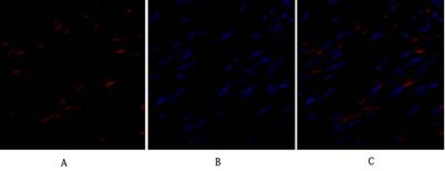
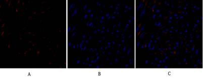
Immunofluorescence analysis of human-uterus tissue. 1,TIMP-1 Polyclonal Antibody(red) was diluted at 1:200(4°C,overnight). 2, Cy3 labled Secondary antibody was diluted at 1:300(room temperature, 50min). 3, Picture B: DAPI(blue) 10min. Picture A:Target.

Immunofluorescence analysis of human-uterus tissue. 1,TIMP-1 Polyclonal Antibody(red) was diluted at 1:200(4°C,overnight). 2, Cy3 labled Secondary antibody was diluted at 1:300(room temperature, 50min). 3, Picture B: DAPI(blue) 10min. Picture A:Target. Picture B: DAPI. Picture C: merge of A+B

No documents available.