TMEM119 Rabbit mAb

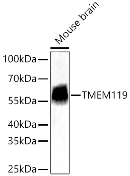
Western blot analysis of lysates from Mouse brain using TMEM119 Rabbit mAb
 at 1:12000 dilution incubated overnight at 4℃.
Secondary antibody: HRP-conjugated Goat anti-Rabbit IgG (H+L)  at 1:10000
dilution.
Lysates/proteins: 25 μg per lane.
Blocking buffer: 3% nonfat dry milk in TBST.
Detection: ECL West Pico Plus.
Exposure time: 45s.

Western blot analysis of lysates from Mouse brain using TMEM119 Rabbit mAb at 1:12000 dilution incubated overnight at 4℃. Secondary antibody: HRP-conjugated Goat anti-Rabbit IgG (H+L) at 1:10000 dilution. Lysates/proteins: 25 μg per lane. Blocking buffer: 3% nonfat dry milk in TBST. Detection: ECL West Pico Plus. Exposure time: 45s.

Catalog Number:MAB27143
Conjugate:Unconjugated
Size:100 ug
Host:Rabbit
Isotype:IgG
Immunogen:Recombinant protein (or fragment).This information is considered to be commercially sensitive.
Reactivity:Human,Mouse,Rat
Applications:WB 1:2000 - 1:24000; IF-F 1:200 - 1:400; IF-P 1:500 - 1:2000; IHC-P 1:5000 - 1:20000; mIHC 1:2000 - 1:8000; ELISA Recommended starting concentration is 1 μg/mL. Please optimize the concentration based on your specific assay requirements.
Molecular Weight:56 kDa
Purification:Affinity purification
Form:Liquid
Synonyms:OBIF

Involved in positive regulation of bone mineralization; positive regulation of osteoblast differentiation; and positive regulation of osteoblast proliferation. Located in plasma membrane. Note:This product is a modified chimeric antibody. For multiplex staining of multiple targets, we recommend using Fc-specific secondary antibodies.

Applications

WB 1:2000 - 1:24000; IF-F 1:200 - 1:400; IF-P 1:500 - 1:2000; IHC-P 1:5000 - 1:20000; mIHC 1:2000 - 1:8000; ELISA Recommended starting concentration is 1 μg/mL. Please optimize the concentration based on your specific assay requirements.

Immunogen

Recombinant protein (or fragment).This information is considered to be commercially sensitive.
Target Background

Involved in positive regulation of bone mineralization; positive regulation of osteoblast differentiation; and positive regulation of osteoblast proliferation. Located in plasma membrane. Note:This product is a modified chimeric antibody. For multiplex staining of multiple targets, we recommend using Fc-specific secondary antibodies.

Synonyms:OBIF

Storage

Store at -20℃. Avoid freeze / thaw cycles.

Buffer

PBS with 0.09% Sodium azide,0.05% BSA,50% glycerol,pH7.3.

Western blot analysis of lysates from Mouse brain using TMEM119 Rabbit mAb at 1:12000 dilution incubated overnight at 4℃. Secondary antibody: HRP-conjugated Goat anti-Rabbit IgG (H+L) at 1:10000 dilution. Lysates/proteins: 25 μg per lane. Blocking buffer: 3% nonfat dry milk in TBST. Detection: ECL West Pico Plus. Exposure time: 45s.

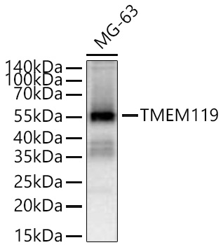
Western blot analysis of lysates from MG-63 cells using TMEM119 Rabbit mAb at 1:2000 dilution incubated overnight at 4℃. Secondary antibody: HRP-conjugated Goat anti-Rabbit IgG (H+L) at 1:10000 dilution. Lysates/proteins: 25 μg per lane. Blocking buffer: 3% non fat dry milk in TBST. Detection: ECL West Pico Plus. Exposure time: 60s.

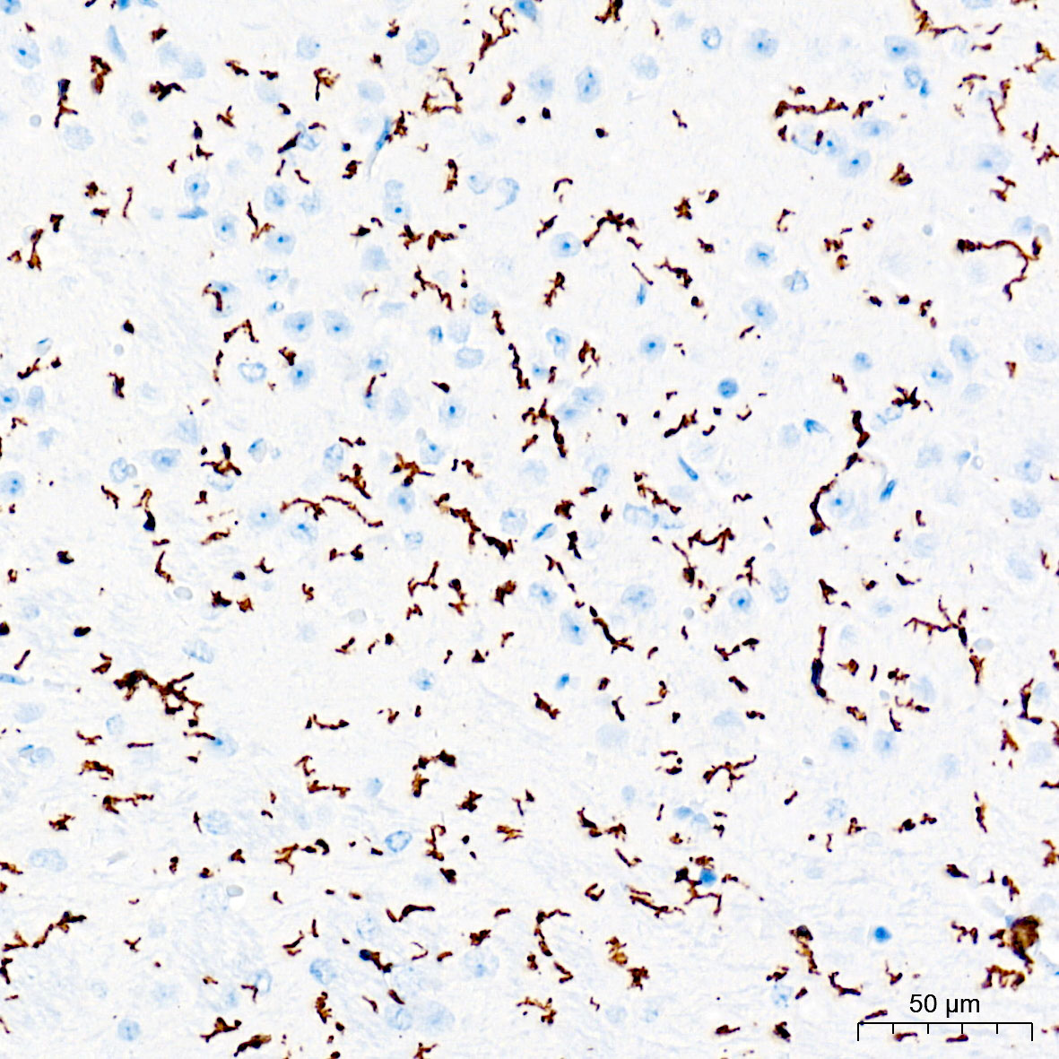
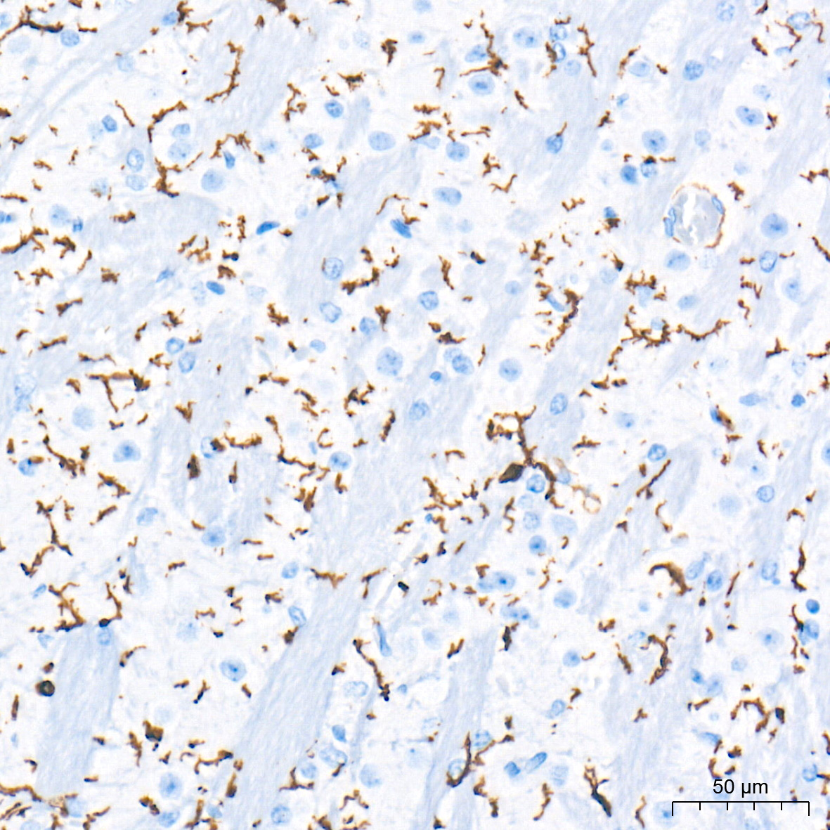
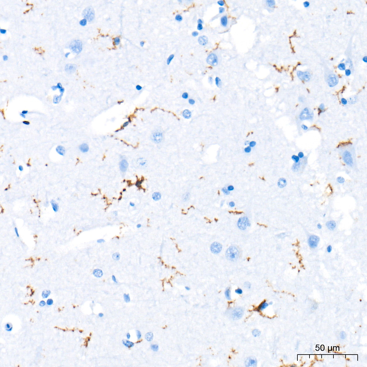
Immunohistochemistry analysis of paraffin-embedded Mouse brain tissue using TMEM119 Rabbit mAb at a dilution of 1:6000 (40x lens). High pressure antigen retrieval performed with 0.01M Tris-EDTA Buffer (pH 9.0) prior to IHC staining.

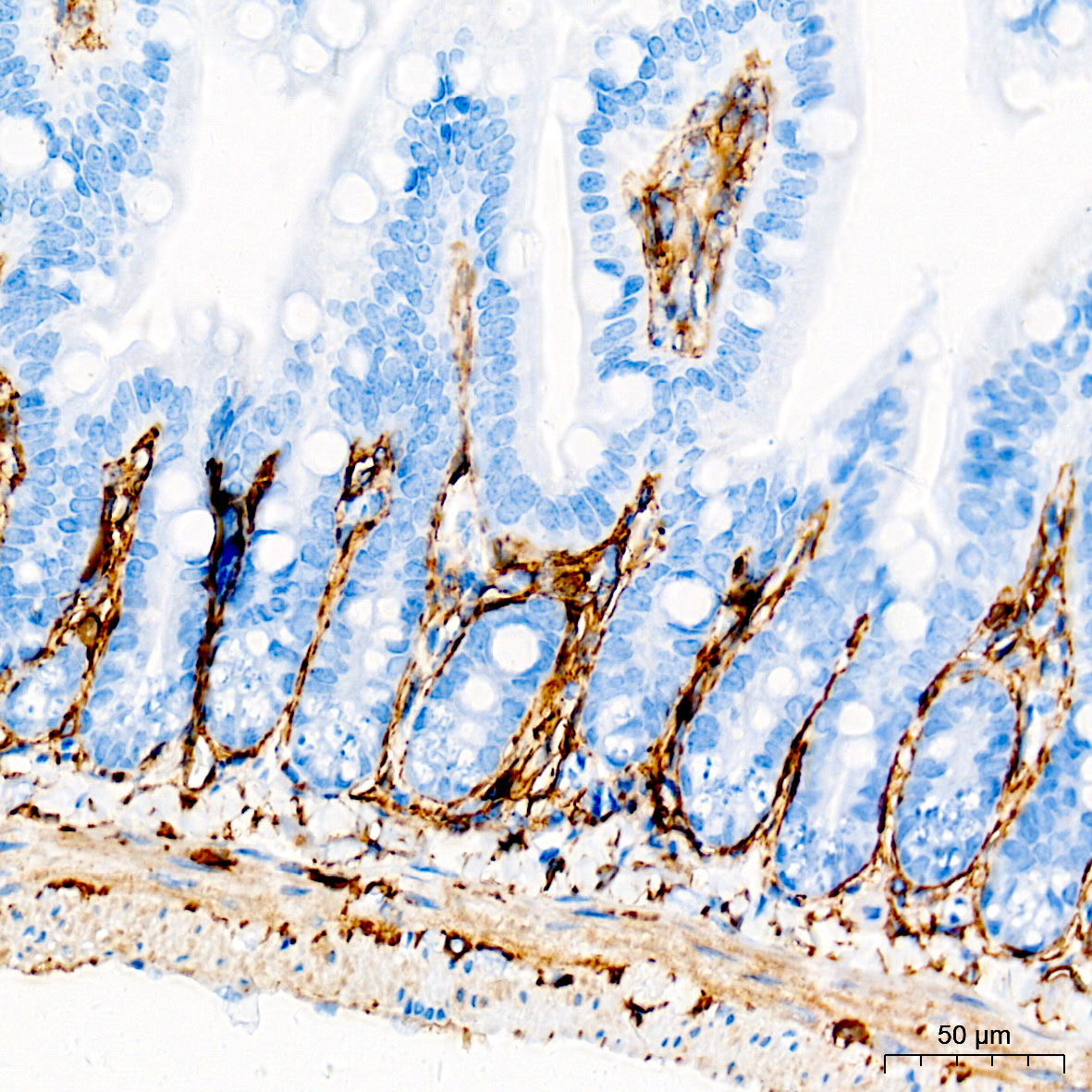
Immunohistochemistry analysis of paraffin-embedded Mouse intestine tissue using TMEM119 Rabbit mAb at a dilution of 1:6000 (40x lens). High pressure antigen retrieval performed with 0.01M Tris-EDTA Buffer (pH 9.0) prior to IHC staining.

Immunohistochemistry analysis of paraffin-embedded Mouse brain tissue using TMEM119 Rabbit mAb at a dilution of 1:6000 (40x lens). High pressure antigen retrieval performed with 0.01M Tris-EDTA Buffer (pH 9.0) prior to IHC staining.

Immunohistochemistry analysis of paraffin-embedded Mouse brain tissue using TMEM119 Rabbit mAb at a dilution of 1:6000 (40x lens). High pressure antigen retrieval performed with 0.01M Tris-EDTA Buffer (pH 9.0) prior to IHC staining.

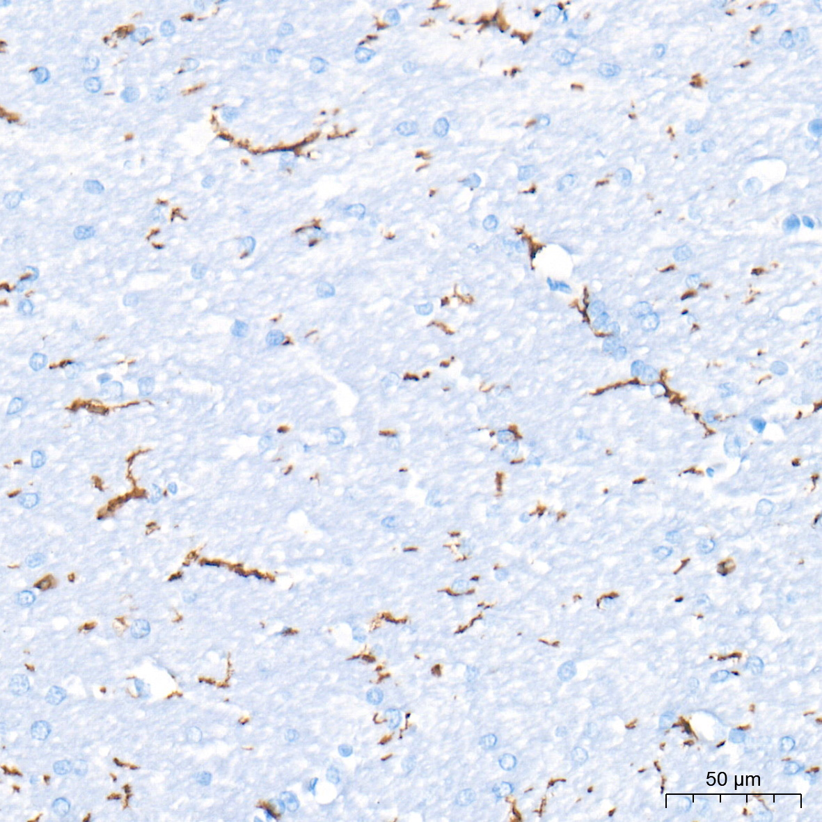
Immunohistochemistry analysis of paraffin-embedded Rat brain tissue using TMEM119 Rabbit mAb at a dilution of 1:6000 (40x lens). High pressure antigen retrieval performed with 0.01M Tris-EDTA Buffer (pH 9.0) prior to IHC staining.

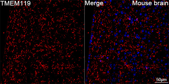
Confocal imaging of paraffinembedded Mouse brain tissue using TMEM119 Rabbit mAb (dilution 1:500) followed by a further incubation with Cy3 Goat Anti-Rabbit IgG (H+L) (dilution 1:500) (Red). DAPI was used for nuclear staining (Blue). High pressure antigen retrieval performed with 0.01M Citrate Buffer (pH 6.0) prior to IF staining. Objective: 40x.

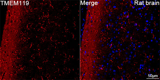
Confocal imaging of paraffinembedded Rat brain tissue using TMEM119 Rabbit mAb dilution 1:1000) followed by a further incubation with Cy3 Goat Anti-Rabbit IgG (H+L) dilution 1:500) (Red). DAPI was used for nuclear staining (Blue). High pressure antigen retrieval performed with 0.01M Citrate Buffer (pH 6.0) prior to IF staining. Objective: 40x.

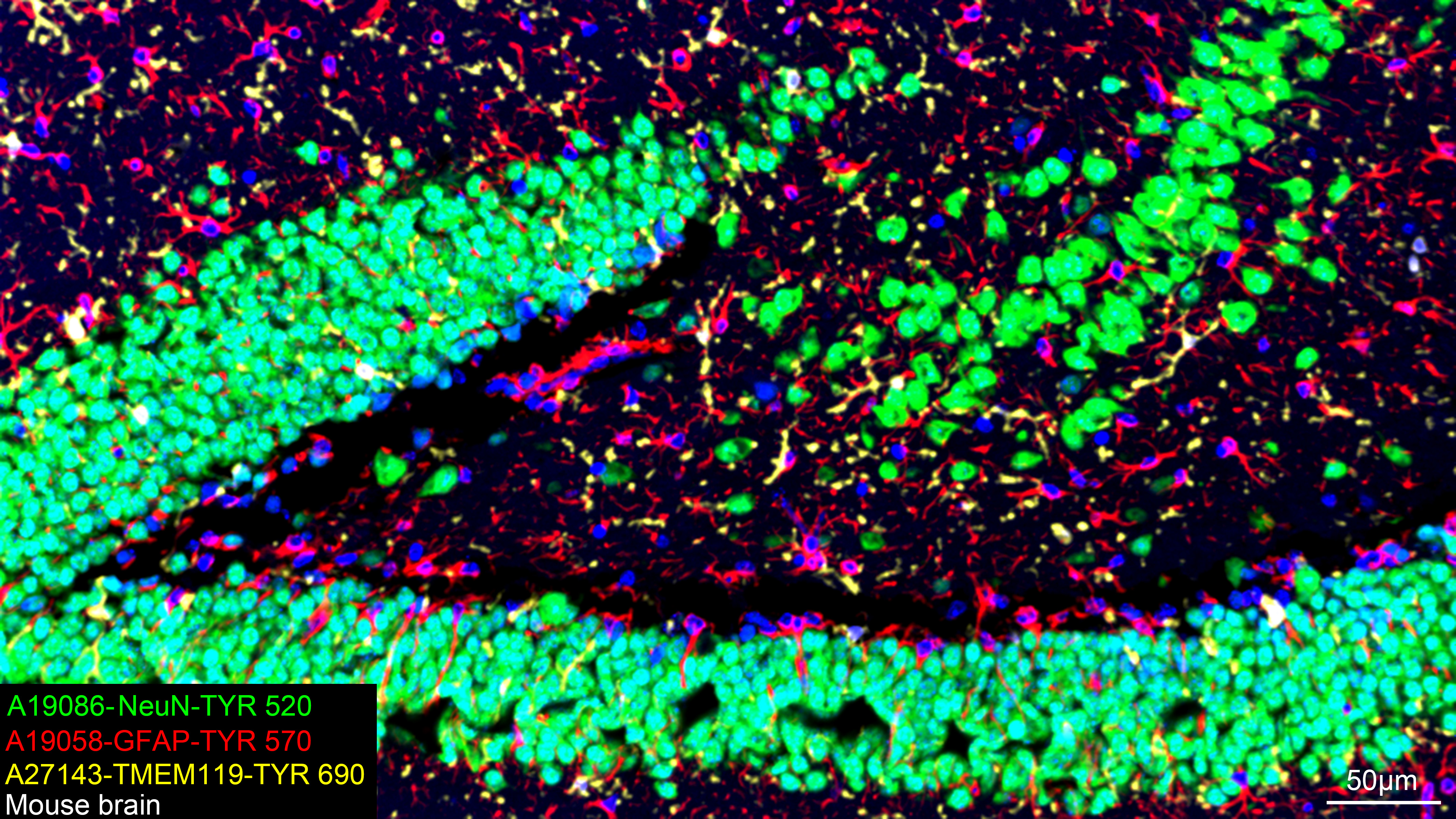
The multiplex IHC analysis on paraffin-embedded Mouse brain tissue using the following specific primary antibodies and tyramide signal amplification (TSA) reagents: NeuN Rabbit mAb (1:2000) with TSA-TYR-520 (Green), GFAP Rabbit mAb (1:500) with TSA-TYR-570 (Red), and TMEM119 Rabbit mAb 1:600) with TSA-TYR-690 (Yellow). DAPI (Blue) was used for nuclear staining. Prior to multiplex IHC staining, high-pressure antigen retrieval was performed using 0.01M citrate buffer at pH 6.0. The analysis was completed using a 20x objective lens.

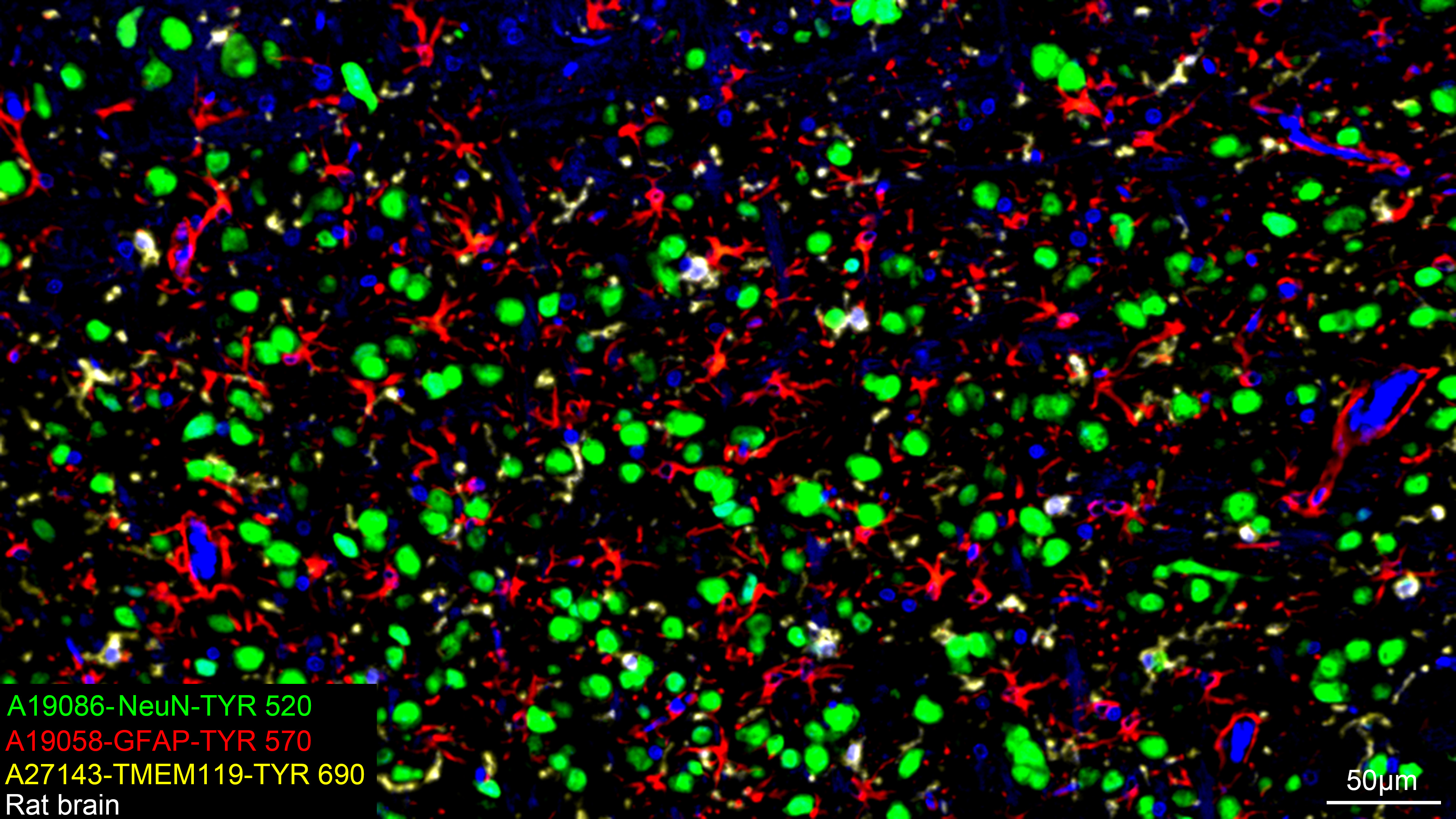
The multiplex IHC analysis on paraffin-embedded Rat brain tissue using the following specific primary antibodies and tyramide signal amplification (TSA) reagents NeuN Rabbit mAb (1:2000) with TSA-TYR-520 (Green), GFAP Rabbit mAb (1:500) with TSA-TYR-570 (Red), and TMEM119 Rabbit mAb 1:600) with TSA-TYR-690 (Yellow). DAPI (Blue) was used for nuclear staining. Prior to multiplex IHC staining, high-pressure antigen retrieval was performed using 0.01M citrate buffer at pH 6.0. The analysis was completed using a 20x objective lens.

No documents available.