TMEM119 Rabbit Polyclonal Antibody

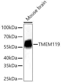
Western blot analysis of lysates from Mouse brain using TMEM119 Rabbit  at 1:12000 dilution incubated overnight at 4℃.
Secondary antibody: HRP-conjugated Goat anti-Rabbit IgG (H+L)  at 1:10000dilution.
Lysates/proteins: 25 μg per lane.
Blocking buffer: 3% nonfat dry milk in TBST.
Detection: ECL West Pico Plus.
Exposure time: 45s.

Western blot analysis of lysates from Mouse brain using TMEM119 Rabbit  at 1:12000 dilution incubated overnight at 4℃. Secondary antibody: HRP-conjugated Goat anti-Rabbit IgG (H+L)  at 1:10000dilution. Lysates/proteins: 25 μg per lane. Blocking buffer: 3% nonfat dry milk in TBST. Detection: ECL West Pico Plus. Exposure time: 45s.

Catalog Number:AB-84816
Conjugate:Unconjugated
Size:100 ug
Concentration:1mg/ml
Host:Rabbit
Isotype:IgG
Clone:POLY
Immunogen:Recombinant protein of human TMEM119.
Reactivity:Mouse, Human
Applications:Western Blot:    1:6000 - 1:24000 Immunohistochemistry (paraffin-embedded tissues):  1:2000 - 1:8000 Immunofluorescence:  1:500 - 1:2000 Immunocytochemistry:  1:500 - 1:2000 ELISA Recommended starting concentration is 1 μg/mL.
Molecular Weight:56kDa
Purification:Affinity Purified
Form:Liquid
Synonyms:OBIF

Applications

Western Blot:    1:6000 - 1:24000 Immunohistochemistry (paraffin-embedded tissues):  1:2000 - 1:8000 Immunofluorescence:  1:500 - 1:2000 Immunocytochemistry:  1:500 - 1:2000 ELISA Recommended starting concentration is 1 μg/mL.

Immunogen

Recombinant protein of human TMEM119.
Target Background

Involved in positive regulation of bone mineralization; positive regulation of osteoblast differentiation; and positive regulation of osteoblast proliferation. Located in plasma membrane.

Synonyms:OBIF

Storage

Store at -20℃. Avoid freeze / thaw cycles.

Buffer

PBS with 0.09% Sodium azide,0.05% BSA,50% glycerol,pH7.3.

Western blot analysis of lysates from Mouse brain using TMEM119 Rabbit  at 1:12000 dilution incubated overnight at 4℃. Secondary antibody: HRP-conjugated Goat anti-Rabbit IgG (H+L)  at 1:10000dilution. Lysates/proteins: 25 μg per lane. Blocking buffer: 3% nonfat dry milk in TBST. Detection: ECL West Pico Plus. Exposure time: 45s.

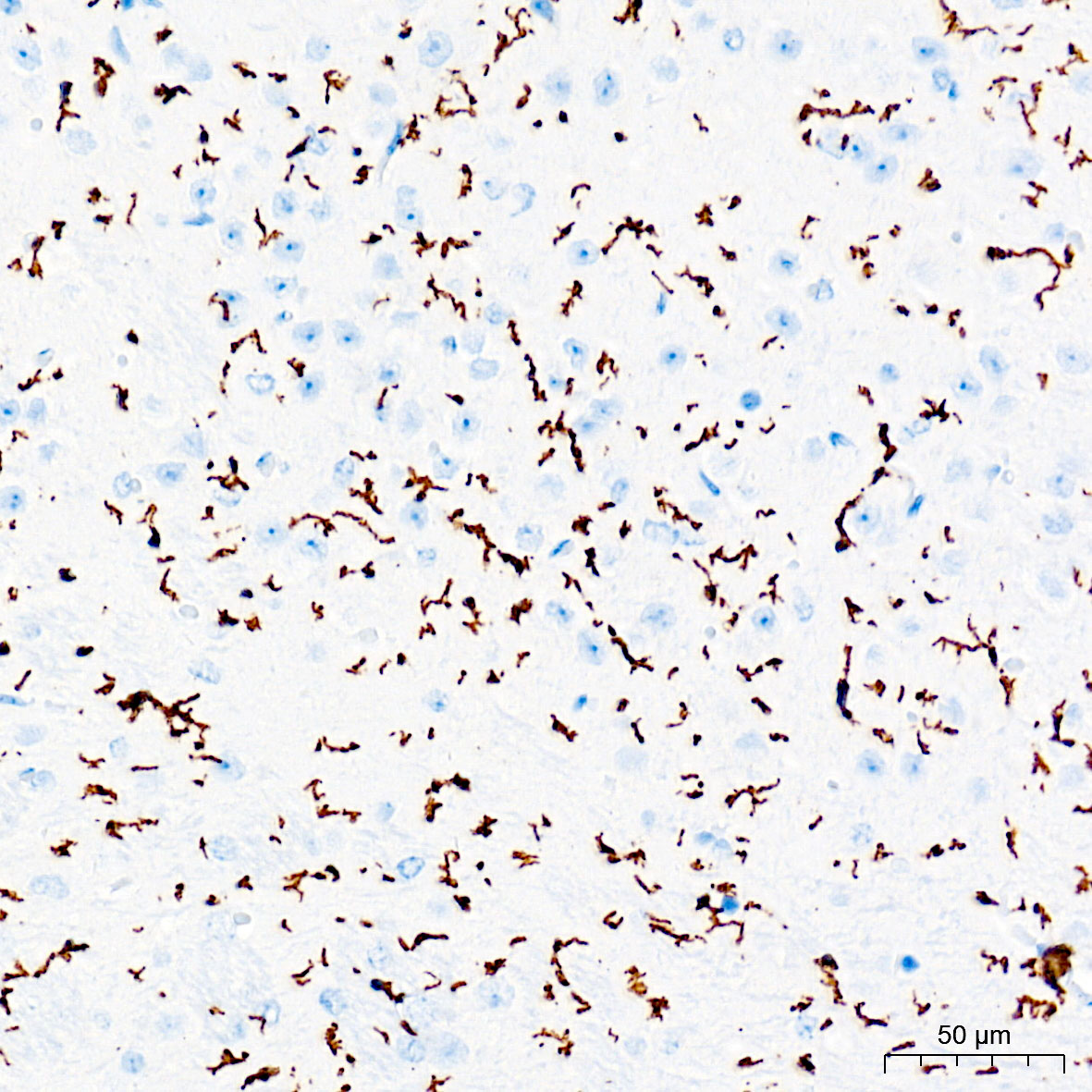
Immunohistochemistry analysis of paraffin-embedded Mouse brain tissue using TMEM119 Rabbit at a dilution of 1:6000 (40xlens). High pressure antigen retrieval performed with 0.01M Tris-EDTA Buffer (pH 9.0) prior to IHC staining.

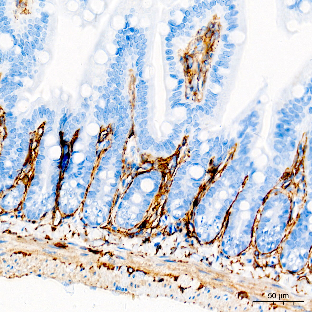
Immunohistochemistry analysis of paraffin-embedded Mouse intestine tissue using TMEM119 Rabbit at a dilution of 1:6000 (40xlens). High pressure antigen retrieval performed with 0.01M Tris-EDTA Buffer (pH 9.0) prior to IHC staining.

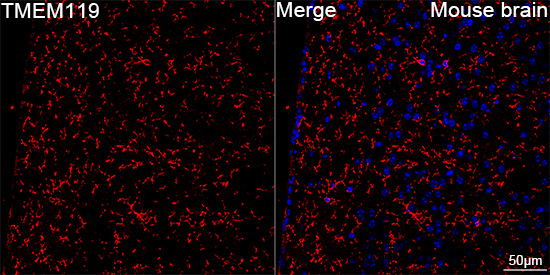
Confocal imaging of paraffinembedded Mouse brain tissue usingTMEM119 Rabbitdilution 1:500) followed by a further incubation with Cy3 Goat Anti-Rabbit IgG (H+L) (AS007, dilution 1:500) (Red). DAPI was used for nuclear staining (Blue). High pressure antigen retrieval performed with 0.01M Citrate Buffer (pH 6.0) prior to IF staining. Objective: 40x.

No documents available.