TNF alpha

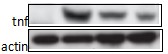
Western blot analysis of extracts of various cell lines, using TNF-α antibody at 1:1000 dilution.
Secondary antibody: HRP Goat Anti-Rabbit IgG (H+L) at 1:10000 dilution.
Lysates/proteins: 25ug per lane.
Blocking buffer: 3% nonfat dry milk in TBST.Detection: ECL West Pico Plus.
Exposure time: 30s.

Western blot analysis of extracts of various cell lines, using TNF-α antibody at 1:1000 dilution. Secondary antibody: HRP Goat Anti-Rabbit IgG (H+L) at 1:10000 dilution. Lysates/proteins: 25ug per lane. Blocking buffer: 3% nonfat dry milk in TBST.Detection: ECL West Pico Plus. Exposure time: 30s.

Catalog Number:MAB-94636
Conjugate:Unconjugated
Size:100 ug
Concentration:1mg/ml
Host:Rb
Isotype:IgG
Clone:TNF-3
Immunogen:Recombinant fusion protein containing a sequence corresponding to amino acids 1-233 of human TNF.
Reactivity:Hu, Ms, Rt
Applications:Western Blot: 1:1000 - 1:2000 Immunohistochemistry: 1:50 - 1:200 Immunofluorescence: 1:50 - 1:200
Molecular Weight:17kDa
Purification:Aff. Pur.
Form:Liquid
Synonyms:TNF;DIF;TNF-alpha;TNFA;TNFSF2;TNLG1F;TNF alpha

Applications

Western Blot: 1:1000 - 1:2000 Immunohistochemistry: 1:50 - 1:200 Immunofluorescence: 1:50 - 1:200

Immunogen

Recombinant fusion protein containing a sequence corresponding to amino acids 1-233 of human TNF.
Target Background

This gene encodes a multifunctional proinflammatory cytokine that belongs to the tumor necrosis factor (TNF) superfamily. This cytokine is mainly secreted by macrophages. It can bind to, and thus functions through its receptors TNFRSF1A/TNFR1 and TNFRSF1B/TNFBR. This cytokine is involved in the regulation of a wide spectrum of biological processes including cell proliferation, differentiation, apoptosis, lipid metabolism, and coagulation. This cytokine has been implicated in a variety of diseases,including autoimmune diseases, insulin resistance, and cancer. Knockout studies in mice also suggested the neuroprotective function of this cytokine.

Synonyms:TNF;DIF;TNF-alpha;TNFA;TNFSF2;TNLG1F;TNF alpha

Storage

Store at -20℃. Avoid freeze / thaw cycles.

Buffer

PBS with 0.02% sodium azide,50% glycerol, pH7.3.

Western blot analysis of extracts of various cell lines, using TNF-α antibody at 1:1000 dilution. Secondary antibody: HRP Goat Anti-Rabbit IgG (H+L) at 1:10000 dilution. Lysates/proteins: 25ug per lane. Blocking buffer: 3% nonfat dry milk in TBST.Detection: ECL West Pico Plus. Exposure time: 30s.

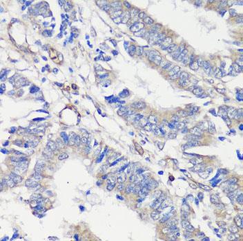
Immunohistochemistry of paraffin embedded Human colon carcinoma using TNF-α antibody at dilution of 1:100 (40x lens).

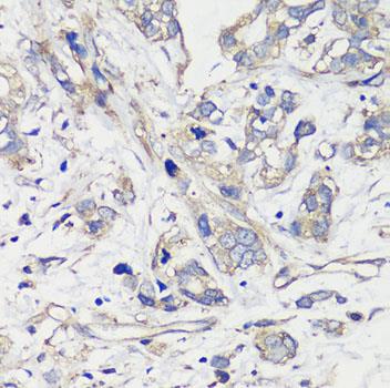
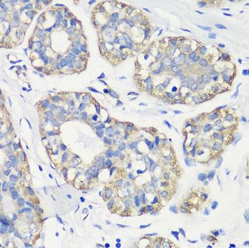
Immunohistochemistry of paraffin embedded Human breast cancer using TNF-α antibody at dilution of 1:100 (40x lens).

Immunohistochemistry of paraffin Embedded Human gastric cancer using TNF-α antibody at dilution of 1:100 (40x lens).

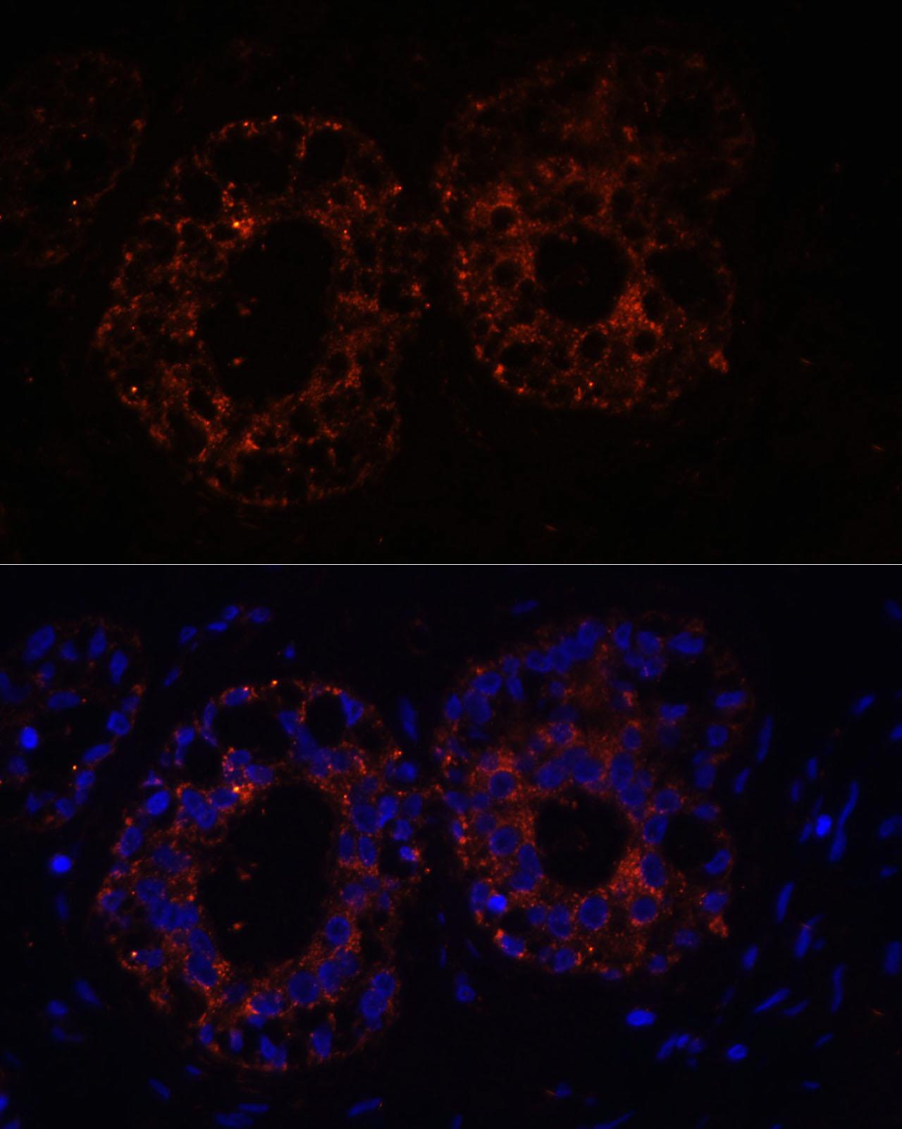
Immunofluorescence analysis of Human mammary cancer cells using TNF-α antibody at dilution of 1:100. Blue: DAPI for nuclear staining.

Review: Review for TNF-α Rabbit mAb Experiment Type Western blot (WB)

No documents available.