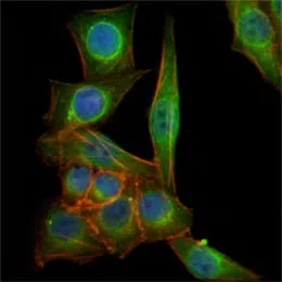
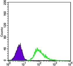
TUBB3 Mouse Monoclonal Antibody
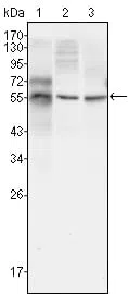
Western blot analysis using TUBB3 mouse mAb against HepG2 (1), A549 (2) and Hela (3) cell lysate.
| Catalog Number: | MABN80763 |
| Conjugate: | Unconjugated |
| Size: | 100ul |
| Concentration: | 1mg/ml |
| Host: | Mouse |
| Isotype: | Mouse IgG1 |
| Clone: | Monoclonal |
| Immunogen: | Purified recombinant fragment of human TUBB3 expressed in E. Coli. |
| Reactivity: | Human,Mouse,Rat |
| Applications: | WB 1:500-1:2000,ICC 1:200-1:1000,ELISA 1:5000-1:20000,FC 1:200-1:400 |
| Molecular Weight: | 55kDa |
| Purification: | Affinity purification |
| Form: | liquid |
| Synonyms: | tubulin, beta 3; MC1R; TUBB4 |
Tubulin, beta 3, also known as TUBB3. Tubulin is the major constituent of microtubules. It binds two moles of GTP, one at an exchangeable site on the beta chain and one at a non exchangeable site on the alpha-chain. Tubulin is a highly conserved protein with a molecular weight of ~50 kD. Microtubules play key roles in chromosome segregation in mitosis, intracellular transport, ciliary and flagellar bending, and structural support of the cytoskeleton. The two main tubulin isoforms, a- and
Applications
WB 1:500-1:2000,ICC 1:200-1:1000,ELISA 1:5000-1:20000,FC 1:200-1:400
Immunogen
Purified recombinant fragment of human TUBB3 expressed in E. Coli.