Tubulin beta Mouse Monoclonal Antibody

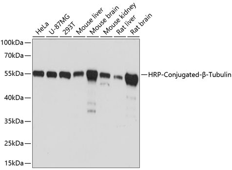
Western blot analysis of extracts of various cell lines, using β-Tubulin antibody HRP conj. at 1:5,000 dilution.
Secondary antibody: HRP Goat Anti-Mouse IgG (H+L) at 1:10,000 dilution.
Lysates/proteins: 25ug per lane.
Blocking buffer: 3% nonfat dry milk in TBST.
Detection: ECL West Pico Plus Kit.
Exposure time: 1s.

Western blot analysis of extracts of various cell lines, using β-Tubulin antibody HRP conj. at 1:5,000 dilution. Secondary antibody: HRP Goat Anti-Mouse IgG (H+L) at 1:10,000 dilution. Lysates/proteins: 25ug per lane. Blocking buffer: 3% nonfat dry milk in TBST. Detection: ECL West Pico Plus Kit. Exposure time: 1s.

Catalog Number:MAB-80143
Conjugate:Unconjugated
Size:100 ug
Concentration:1mg/ml
Host:Ms
Isotype:IgG1
Clone:TUB2
Immunogen:Recombinant protein of human β-Tubulin
Reactivity:Hu, Ms, Rt
Applications:Western Blot: 1:5000 - 1:10000Immunohistochemistry (paraffin-embedded tissues): 1:50 - 1:200Immunofluorescence: 1:50 - 1:200 Immunocytochemistry: 1:50 - 1:200Immunoprecipitation: 1:50- 1:200
Molecular Weight:50kDa
Purification:Aff. Pur.
Form:Liquid
Synonyms:M40; TUBB1; TUBB5; CDCBM6; CSCSC1; OK/SW-cl.56

Applications

Western Blot: 1:5000 - 1:10000Immunohistochemistry (paraffin-embedded tissues): 1:50 - 1:200Immunofluorescence: 1:50 - 1:200 Immunocytochemistry: 1:50 - 1:200Immunoprecipitation: 1:50- 1:200

Immunogen

Recombinant protein of human β-Tubulin
Target Background

This gene encodes a beta tubulin protein. This protein forms a dimer with alpha tubulin and acts as a structural component of microtubules. Mutations in this gene cause cortical dysplasia, complex, with other brain malformations 6. Alternative splicing results in multiple splice variants. There are multiple pseudogenes for this gene on chromosomes 1, 6, 7, 8, 9, and 13.

Synonyms:M40; TUBB1; TUBB5; CDCBM6; CSCSC1; OK/SW-cl.56

Storage

Store at -20℃. Avoid freeze / thaw cycles.

Buffer

PBS with 0.02% sodium azide,1% BSA,50% glycerol,pH7.3.

Western blot analysis of extracts of various cell lines, using β-Tubulin antibody HRP conj. at 1:5,000 dilution. Secondary antibody: HRP Goat Anti-Mouse IgG (H+L) at 1:10,000 dilution. Lysates/proteins: 25ug per lane. Blocking buffer: 3% nonfat dry milk in TBST. Detection: ECL West Pico Plus Kit. Exposure time: 1s.

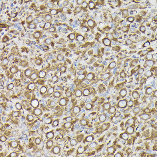
Immunohistochemistry of paraffinembedded rat ovary using β-Tubulin Mouse mAb at dilution of 1:100 (40x lens).Perform high pressure antigen retrieval with 10 mM citrate buffer pH 6.0 before commencing with IHC staining protocol.

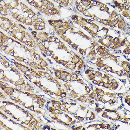
Immunohistochemistry of paraffinembedded rat stomach using β-Tubulin Mouse mAb at dilution of 1:100 (40x lens).Perform high pressure antigen retrieval with 10 mM citrate buffer pH 6.0 before commencing with IHC staining protocol.

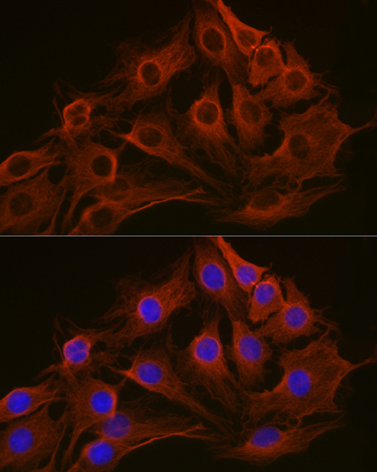
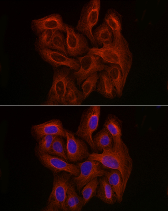
Immunofluorescence analysis of C6 cells using β-Tubulin Mouse mAb at dilution of 1:100 (40x lens). Blue: DAPI for nuclear staining.

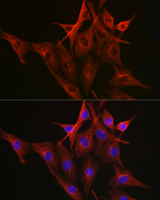
Immunofluorescence analysis of NIH-3T3 cells using β-Tubulin Mouse mAb at dilution of 1:100 (40x lens). Blue: DAPI for nuclear staining.

Immunofluorescence analysis of U-2 OS cells using β-Tubulin Mouse mAb at dilution of 1:100 (40x lens). Blue: DAPI for nuclear staining.

No documents available.