Tubulin beta Rabbit Polyclonal Antibody
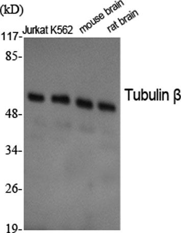
Western Blot analysis of various cells using Tubulin β Polyclonal Antibody diluted at 1:2000. Secondary antibody was diluted at 1:20000

Western Blot analysis of various cells using Tubulin β Polyclonal Antibody diluted at 1:2000. Secondary antibody was diluted at 1:20000

Catalog Number:AB-84819
Conjugate:Unconjugated
Size:100 ug
Concentration:1mg/ml
Host:Rabbit
Isotype:IgG
Clone:POLY
Immunogen:The antiserum was produced against synthesized peptide derived from human Tubulin beta. AA range:401-450.
Reactivity:Human;Rat;Mouse
Applications:Western Blot: 1/500 - 1/2000 Immunohistochemistry (paraffin-embedded tissues):1:100-300 Immunofluorescence:  1:100-300
Molecular Weight:55kD
Purification:The antibody was affinity-purified from rabbit antiserum by affinity-chromatography using epitope-specific immunogen.
Form:Liquid
Synonyms:TUBB3; TUBB4; Tubulin beta-3 chain; Tubulin beta-4 chain; Tubulin beta-III

Applications

Western Blot: 1/500 - 1/2000 Immunohistochemistry (paraffin-embedded tissues):1:100-300 Immunofluorescence:  1:100-300

Immunogen

The antiserum was produced against synthesized peptide derived from human Tubulin beta. AA range:401-450.

Target Background

Beta tubulins are one of two core protein families (alpha and beta tubulins) that heterodimerize and assemble to form microtubules. This protein is primarily expressed in neurons and may be involved in neurogenesis and axon guidance and maintenance. Mutations in this gene are the cause of congenital fibrosis of the extraocular muscles type 3. Alternate splicing results in multiple transcript variants. A pseudogene of this gene is found on chromosome 6.

Synonyms:TUBB3; TUBB4; Tubulin beta-3 chain; Tubulin beta-4 chain; Tubulin beta-III

Storage

Store at -20°C. Avoid repeated freeze-thaw cycles.

Buffer

Liquid in PBS containing 50% glycerol, 0.5% BSA and 0.02% sodium azide.

Western Blot analysis of various cells using Tubulin β Polyclonal Antibody diluted at 1:2000. Secondary antibody was diluted at 1:20000

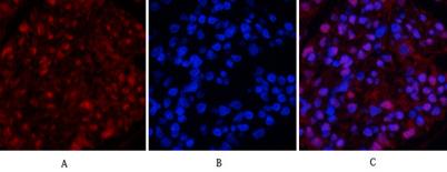
Immunofluorescence analysis of rat-kidney tissue. 1,Tubulin β Polyclonal Antibody(red) was diluted at 1:200(4°C,overnight). 2, Cy3 labled Secondary antibody was diluted at 1:300(room temperature, 50min).3, Picture B: DAPI(blue) 10min. Picture A:Target.

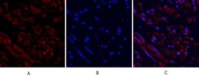
Immunofluorescence analysis of human-uterus tissue. 1,Tubulin β Polyclonal Antibody(red) was diluted at 1:200(4°C,overnight). 2, Cy3 labled Secondary antibody was diluted at 1:300(room temperature, 50min).3, Picture B: DAPI(blue) 10min. Picture A:Target. Picture B: DAPI. Picture C: merge of A+B

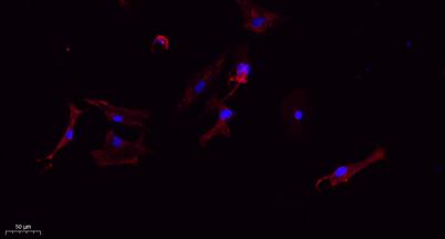
Immunofluorescence analysis of A549. 1,primary Antibody(red) was diluted at 1:200(4°C overnight). 2, Goat Anti Rabbit IgG (H&L) - Alexa Fluor 594 Secondary antibody was diluted at 1:1000(room temperature, 50min).3, Picture B: DAPI(blue) 10min.

No documents available.