Tubulin betaIII
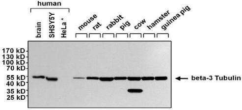
Western Blot analysis using Tubulin Beta III Monoclonal Antibody.
| Catalog Number: | MAB-10288 |
| Conjugate: | Unconjugated |
| Size: | 100 ug |
| Concentration: | 1mg/ml |
| Host: | Ms |
| Isotype: | IgG1 |
| Clone: | TU-20 |
| Immunogen: | Peptide (C) 441-448 coupled to maleimide-activated keyhole limpet hemocyanin via cysteine added to the N-terminus of the neuron-specific peptide. |
| Reactivity: | Hu, Ms, Ch, Bv, Pig, Rt, Ha, Fh |
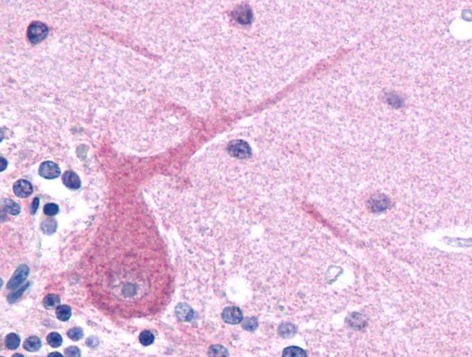
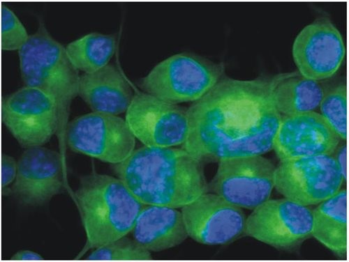
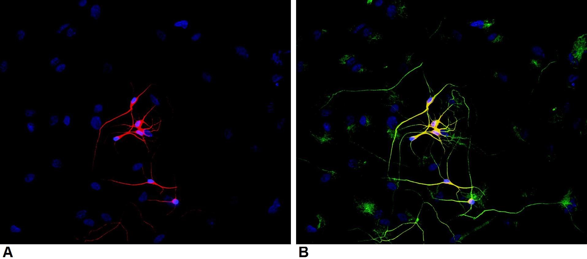
| Applications: | Flow Cytometry Western Blotting Recommended dilution:1-2 µg/ml, 90 min Positive control: Porcine brain lysate Negative control: HPB-ALL human peripheral blood leukemia cell line Sample preparation: Mix lysate with reducing Laemmli SDS-PAGE sample buffer. Application note: Reducing conditions. Immunohistochemistry (paraffin sections) Recommended dilution: 10 µg/ml Staining technique: Standard ABC technique (DAB+) Pretreatment: 0.1% pepsin (trypsin) in 0.1 M HCl; incubation 30 min in RT; or High temperature citrate buffer antigen retrieval Positive tissue: neuronal tissue Immunocytochemistry Positive material: Neuro2a mouse neuroblastoma cell line |
| Purification: | Aff. Pur. |
| Form: | Liquid |
Applications
Flow Cytometry Western Blotting Recommended dilution:1-2 µg/ml, 90 min Positive control: Porcine brain lysate Negative control: HPB-ALL human peripheral blood leukemia cell line Sample preparation: Mix lysate with reducing Laemmli SDS-PAGE sample buffer. Application note: Reducing conditions. Immunohistochemistry (paraffin sections) Recommended dilution: 10 µg/ml Staining technique: Standard ABC technique (DAB+) Pretreatment: 0.1% pepsin (trypsin) in 0.1 M HCl; incubation 30 min in RT; or High temperature citrate buffer antigen retrieval Positive tissue: neuronal tissue Immunocytochemistry Positive material: Neuro2a mouse neuroblastoma cell line
Immunogen
Peptide (C) 441-448 coupled to maleimide-activated keyhole limpet hemocyanin via cysteine added to the N-terminus of the neuron-specific peptide.