Tyrosine Hydroxylase Rabbit mAb

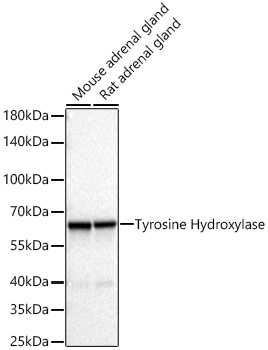
Western blot analysis of various lysates using Tyrosine Hydroxylase Rabbit mAb  at 1:1000 dilution incubated overnight at 4℃.
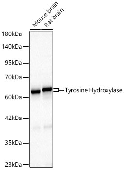
Secondary antibody: HRP-conjugated Goat anti-Rabbit IgG   at 1:10000 dilution.
Lysates / proteins: 25 μg per lane.
Blocking buffer: 3 % nonfat dry milk in TBST.
Detection: ECL Basic Kit .
Exposure time: 10s.

Western blot analysis of various lysates using Tyrosine Hydroxylase Rabbit mAb at 1:1000 dilution incubated overnight at 4℃. Secondary antibody: HRP-conjugated Goat anti-Rabbit IgG at 1:10000 dilution. Lysates / proteins: 25 μg per lane. Blocking buffer: 3 % nonfat dry milk in TBST. Detection: ECL Basic Kit . Exposure time: 10s.

Catalog Number:AB25683
Conjugate:Unconjugated
Size:100 ug
Concentration:1mg/ml
Host:Rabbit
Isotype:IgG
Clone:ARC67477
Immunogen:Recombinant fusion protein containing a sequence corresponding to amino acids 411-528 of human Tyrosine Hydroxylase (NP_954986.2).
Reactivity:Human,Mouse,Rat
Applications:WB,1:1000 - 1:6000 IHC-P,1:3000-1:5000 IF/ICC,1:200 - 1:800 ELISA,Recommended starting concentration is 1 μg/mL. Please optimize the concentration based on your specific assay requirements.
Molecular Weight:62-64kDa
Purification:Affinity purification
Form:liquid

The protein encoded by this gene is involved in the conversion of tyrosine to dopamine. It is the rate-limiting enzyme in the synthesis of catecholamines, hence plays a key role in the physiology of adrenergic neurons. Mutations in this gene have been associated with autosomal recessive Segawa syndrome. Alternatively spliced transcript variants encoding different isoforms have been noted for this gene.

Applications

WB,1:1000 - 1:6000 IHC-P,1:3000-1:5000 IF/ICC,1:200 - 1:800 ELISA,Recommended starting concentration is 1 μg/mL. Please optimize the concentration based on your specific assay requirements.

Immunogen

Recombinant fusion protein containing a sequence corresponding to amino acids 411-528 of human Tyrosine Hydroxylase (NP_954986.2).

Target Background

The protein encoded by this gene is involved in the conversion of tyrosine to dopamine. It is the rate-limiting enzyme in the synthesis of catecholamines, hence plays a key role in the physiology of adrenergic neurons. Mutations in this gene have been associated with autosomal recessive Segawa syndrome. Alternatively spliced transcript variants encoding different isoforms have been noted for this gene.

Storage

Store at -20℃. Avoid freeze / thaw cycles.

Buffer

PBS with 0.09% Sodium azide,0.05% BSA,50% glycerol,pH7.3.

Western blot analysis of various lysates using Tyrosine Hydroxylase Rabbit mAb at 1:1000 dilution incubated overnight at 4℃. Secondary antibody: HRP-conjugated Goat anti-Rabbit IgG at 1:10000 dilution. Lysates / proteins: 25 μg per lane. Blocking buffer: 3 % nonfat dry milk in TBST. Detection: ECL Basic Kit . Exposure time: 10s.

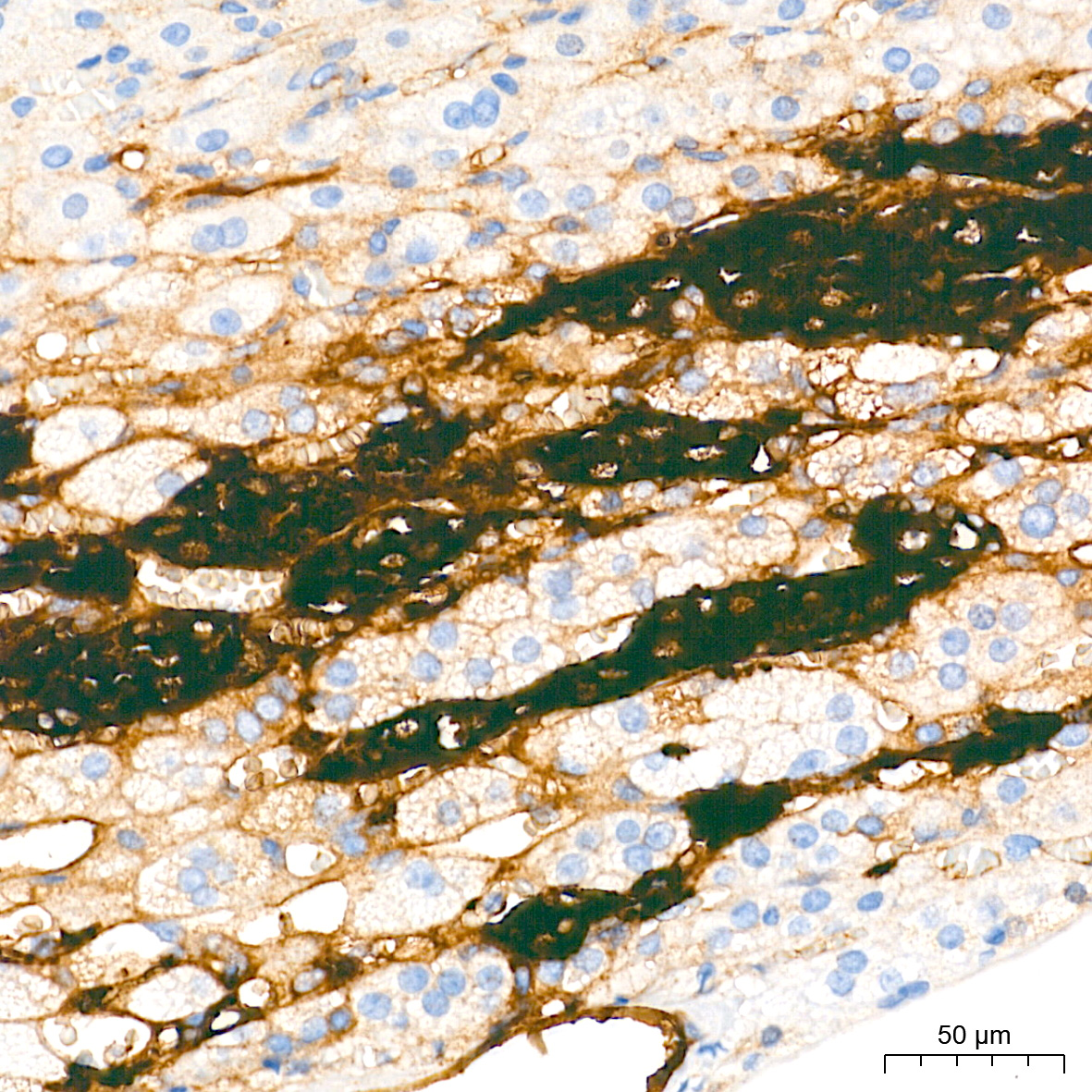
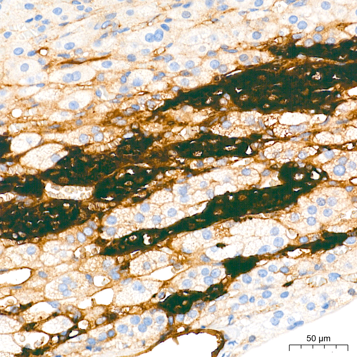
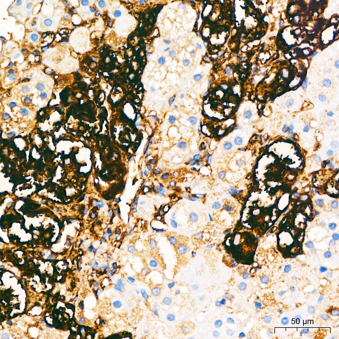
Immunohistochemistry analysis of paraffin-embedded Mouse adrenal gland tissue using Tyrosine Hydroxylase Rabbit mAb at a dilution of 1:4000 . High pressure antigen retrieval performed with 0.01M Citrate Buffer prior to IHC staining.

Immunohistochemistry analysis of paraffin-embedded Human adrenal gland tissue using Tyrosine Hydroxylase Rabbit mAb at a dilution of 1:4000 . High pressure antigen retrieval performed with 0.01M Citrate Buffer prior to IHC staining.

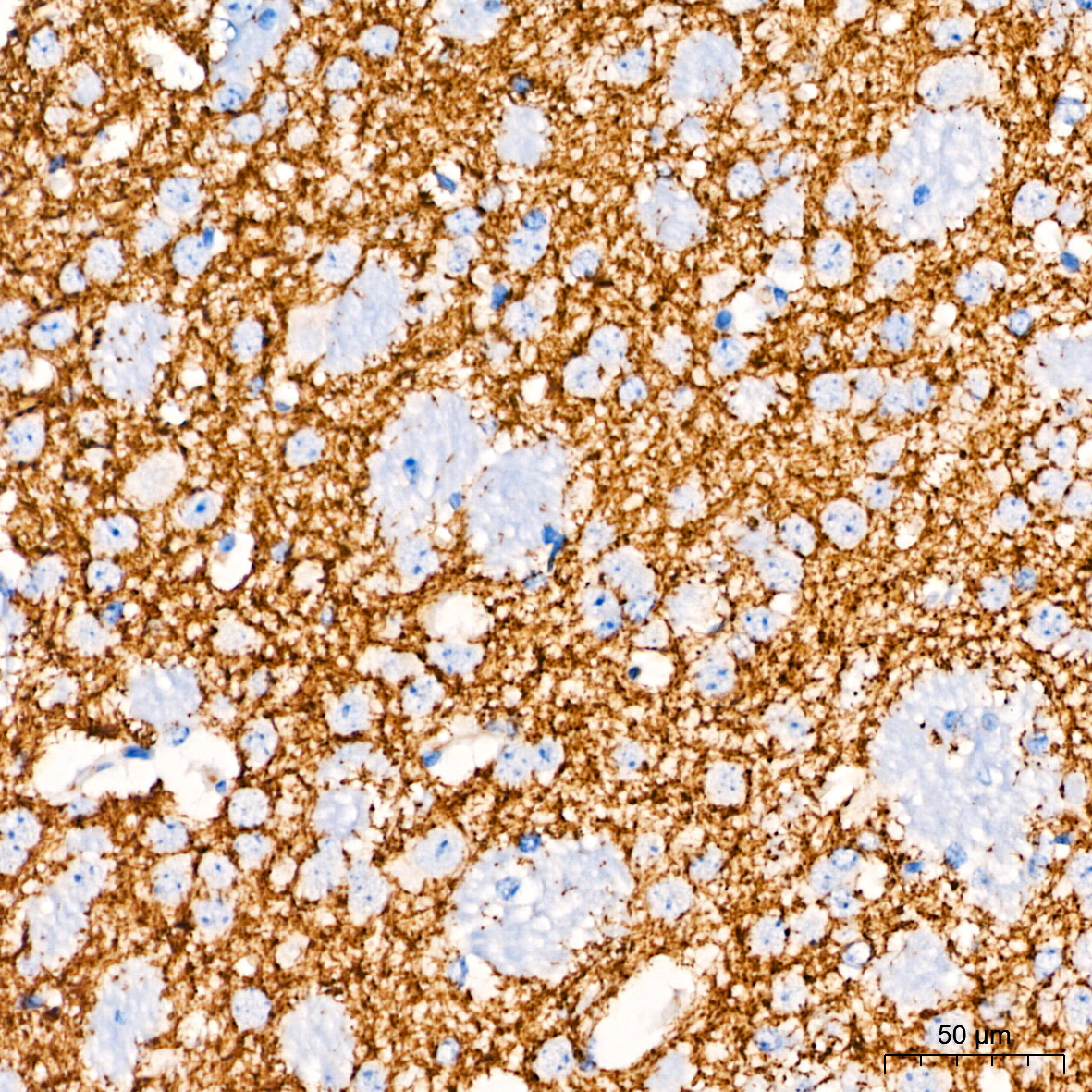
Immunohistochemistry analysis of paraffin-embedded Mouse brain tissue using Tyrosine Hydroxylase Rabbit mAb at a dilution of 1:4000 . High pressure antigen retrieval performed with 0.01M Citrate Buffer prior to IHC staining.

Confocal imaging of 293T cells transfected with TH using Tyrosine Hydroxylase Rabbit mAb followed by a further incubation with Cy3 Goat Anti-Rabbit IgG . DAPI was used for nuclear staining . Objective: 100x.

Confocal imaging of paraffin-embedded Mouse brain tissue using Tyrosine Hydroxylase Rabbit mAb followed by a further incubation with Cy3 Goat Anti-Rabbit IgG . DAPI was used for nuclear staining . Objective: 40x. Perform microwave antigen retrieval with 0.01 M citrate buffer prior to IF staining. Confocal imaging of paraffin-embedded Mouse brain tissue using Tyrosine Hydroxylase Rabbit mAb followed by a further incubation with Cy3 Goat Anti-Rabbit IgG . DAPI was used for nuclear staining . High pressure antigen retrieval performed with 0.01M Citrate Buffer prior to IF staining. Objective: 40x.

No documents available.