β-Actin Rabbit Monoclonal Antibody

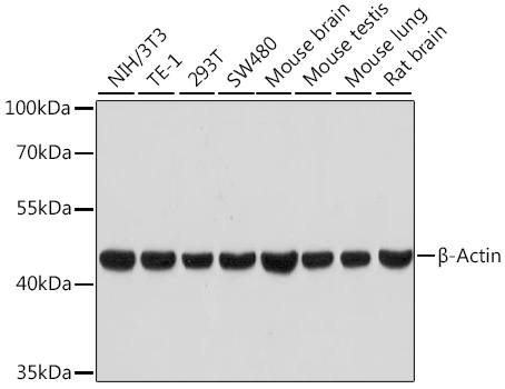
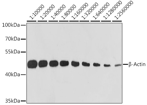
Western blot analysis of various lysates using β-Actin Rabbit mAb at 1:100000 dilution.
Secondary antibody: HRP-conjugated Goat anti-Rabbit IgG (H+L) at 1:10000 dilution.
Lysates/proteins: 25μg per lane.
Blocking buffer: 3% nonfat dry milk in TBST.
Detection: ECL West Pico Plus.
Exposure time: 30s.

Western blot analysis of various lysates using β-Actin Rabbit mAb at 1:100000 dilution. Secondary antibody: HRP-conjugated Goat anti-Rabbit IgG (H+L) at 1:10000 dilution. Lysates/proteins: 25μg per lane. Blocking buffer: 3% nonfat dry milk in TBST. Detection: ECL West Pico Plus. Exposure time: 30s.

Catalog Number:MAB026
Conjugate:Unconjugated
Size:300 ug
Concentration:1mg/ml
Host:Rabbit
Isotype:IgG
Clone:13E5
Immunogen:Recombinant protein.This information is considered to be commercially sensitive.
Reactivity:Human,Mouse,Rat,Chicken,Zebrafish,Pig,Cow
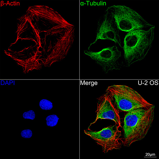
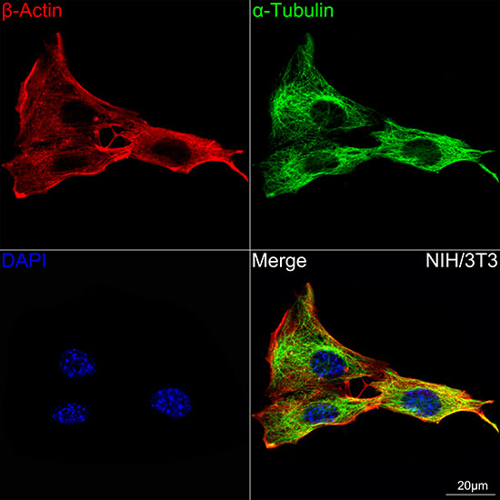
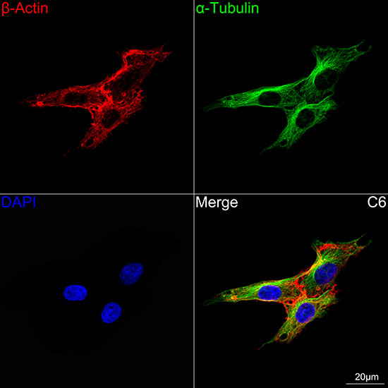
Applications:Western Blot: 1:10000-1:100000 Immunofluorescence: 1:200-1:800 Immunocytochemistry: 1:200-1:800 Immunohistocehmistry(paraffin-embedded tissues): 1:3000 - 1:10000 ELISA
Molecular Weight:42kDa
Purification:Affinity purification
Form:liquid
Synonyms:BRWS1; PS1TP5BP1; β-Actin

This gene encodes one of six different actin proteins. Actins are highly conserved proteins that are involved in cell motility, structure, integrity, and intercellular signaling. The encoded protein is a major constituent of the contractile apparatus and one of the two nonmuscle cytoskeletal actins that are ubiquitously expressed. Mutations in this gene cause Baraitser-Winter syndrome 1, which is characterized by intellectual disability with a distinctive facial appearance in human patients. Numerous pseudogenes of this gene have been identified throughout the human genome. Note:Due to the high antibody titer, it is advisable to be diluted before use. Please dilute 5μL of the antibody solution with 45μL of PBS solution, containing 50% glycerol. The diluted antibody can be stored at -20°C.

Applications

Western Blot: 1:10000-1:100000 Immunofluorescence: 1:200-1:800 Immunocytochemistry: 1:200-1:800 Immunohistocehmistry(paraffin-embedded tissues): 1:3000 - 1:10000 ELISA

Immunogen

Recombinant protein.This information is considered to be commercially sensitive.
Target Background

This gene encodes one of six different actin proteins. Actins are highly conserved proteins that are involved in cell motility, structure, integrity, and intercellular signaling. The encoded protein is a major constituent of the contractile apparatus and one of the two nonmuscle cytoskeletal actins that are ubiquitously expressed. Mutations in this gene cause Baraitser-Winter syndrome 1, which is characterized by intellectual disability with a distinctive facial appearance in human patients. Numerous pseudogenes of this gene have been identified throughout the human genome. Note:Due to the high antibody titer, it is advisable to be diluted before use. Please dilute 5μL of the antibody solution with 45μL of PBS solution, containing 50% glycerol. The diluted antibody can be stored at -20°C.

Synonyms:BRWS1; PS1TP5BP1; β-Actin

Storage

Store at -20℃. Avoid freeze / thaw cycles.

Buffer

PBS containing 50% glycerol and 0.05% BSA, preserved with proclin300 or sodium azide (as specified on the Certificate of Analysis), pH 7.3.

Western blot analysis of various lysates using β-Actin Rabbit mAb at 1:100000 dilution. Secondary antibody: HRP-conjugated Goat anti-Rabbit IgG (H+L) at 1:10000 dilution. Lysates/proteins: 25μg per lane. Blocking buffer: 3% nonfat dry milk in TBST. Detection: ECL West Pico Plus. Exposure time: 30s.

Western blot analysis of lysates from HeLa cells, using β-Actin Rabbit mAb at 1:10000-1:75000 dilution. Secondary antibody: HRP-conjugated Goat anti-Rabbit IgG (H+L) at 1:10000 dilution. Lysates/proteins: 25μg per lane. Blocking buffer: 3% nonfat dry milk in TBST. Detection: ECL West Pico Plus. Exposure time: 10s.

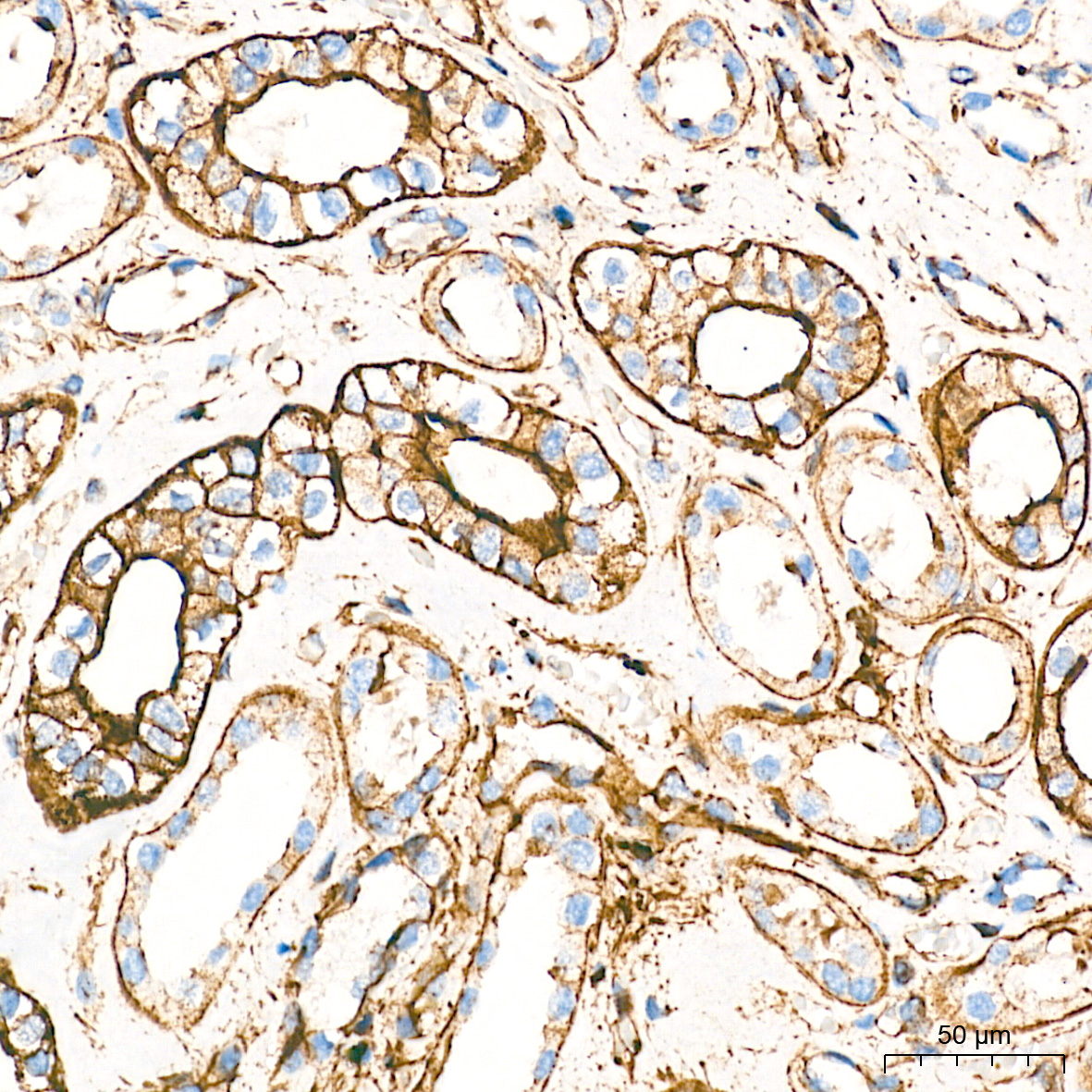
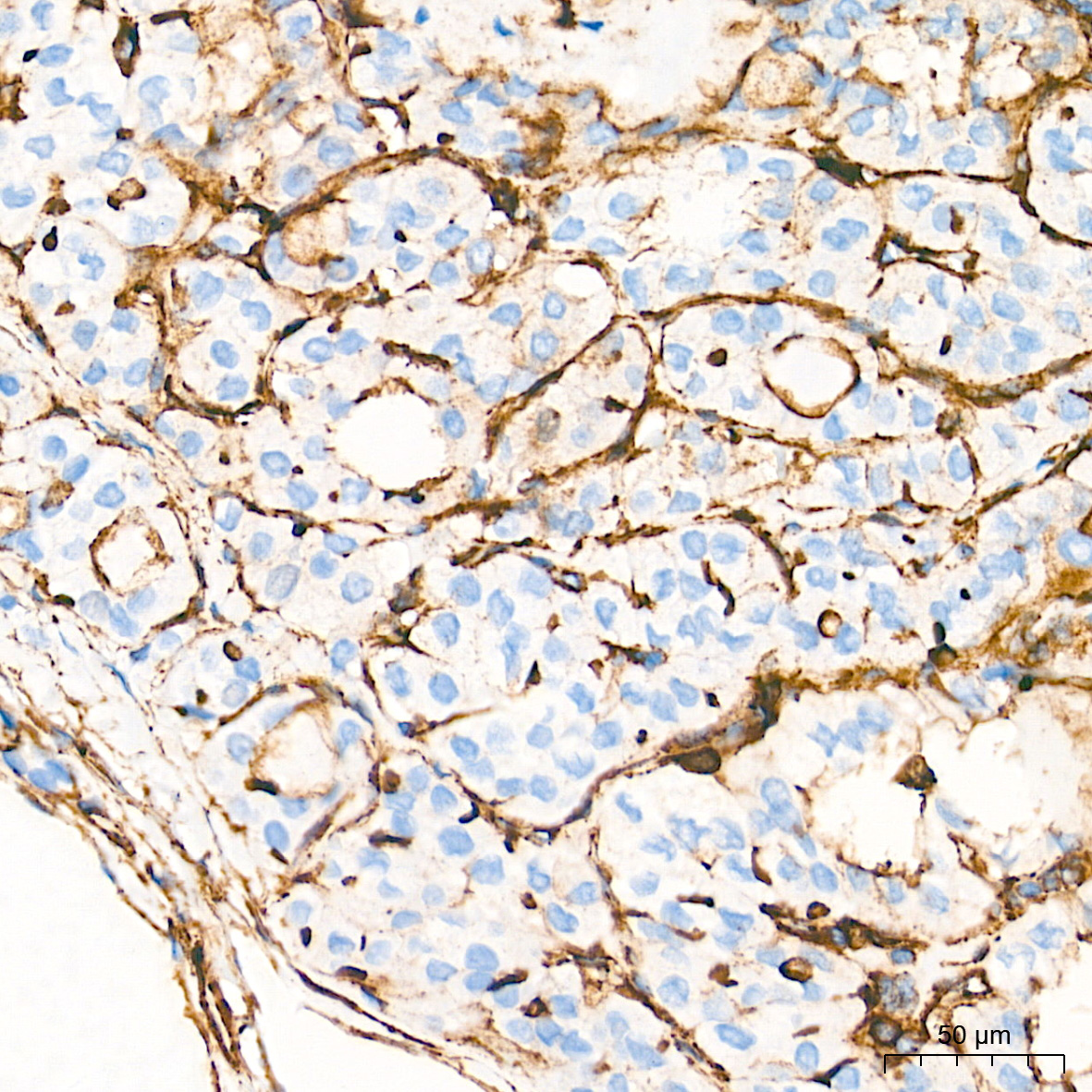
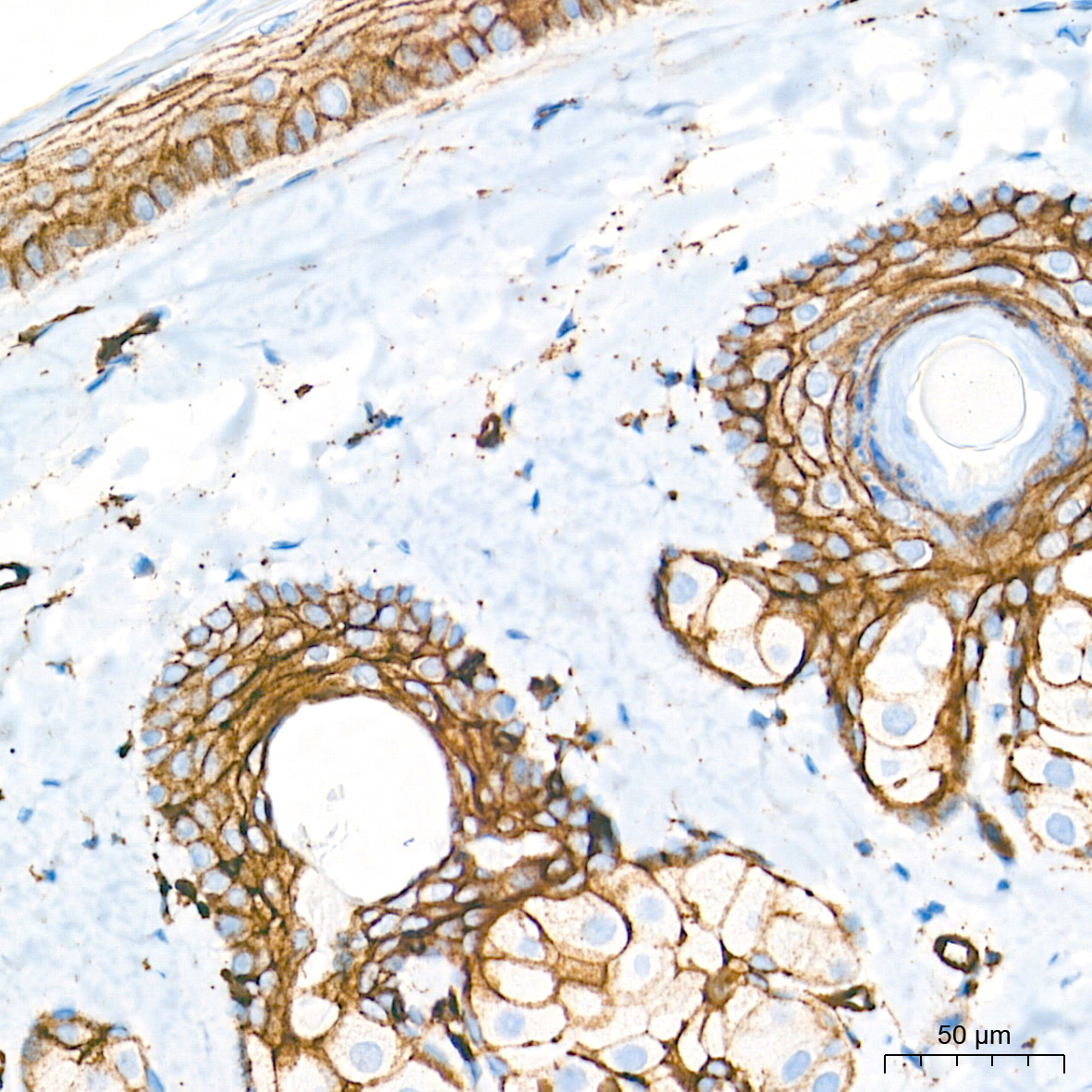
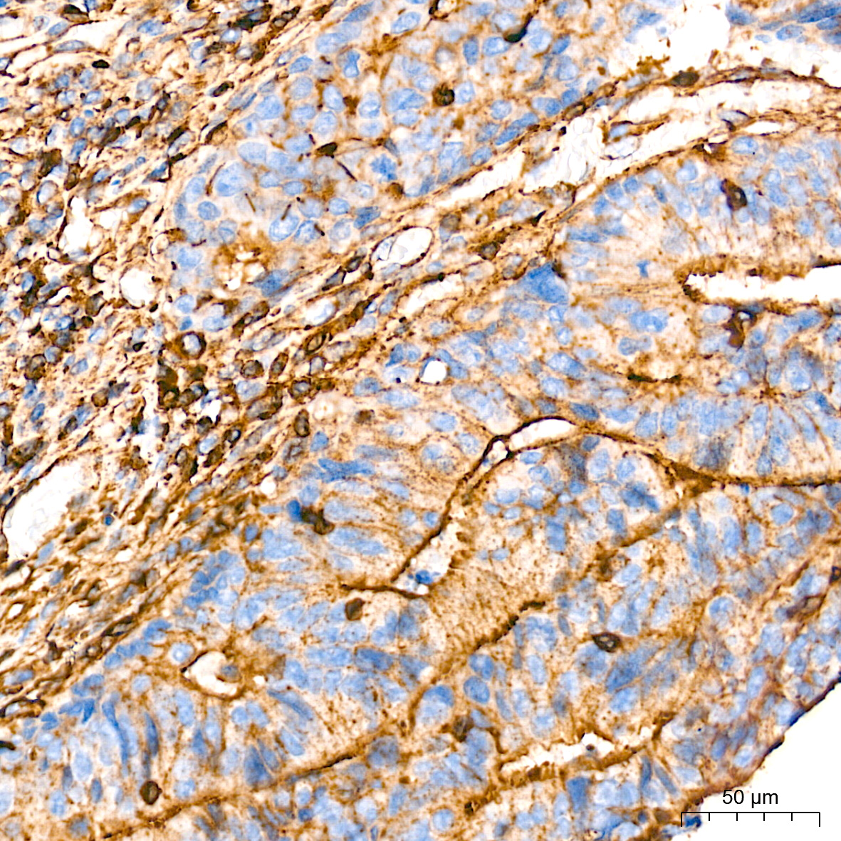
Immunohistochemistry analysis of paraffin-embedded Human kidney tissue using β-Actin Rabbit mAb at a dilution of 1:10000 (40x lens). High pressure antigen retrieval performed with 0.01M Tris-EDTA Buffer (pH 9.0) prior to IHC staining.

No documents available.