β-arrestin1 Rabbit Polyclonal Antibody

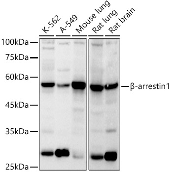
Western blot analysis of various lysates using β-arrestin1 Rabbit pAb  at 1:1000 dilution.
Secondary antibody: HRP-conjugated Goat anti-Rabbit IgG (H+L)  at 1:10000 dilution.
Lysates/proteins: 25μg per lane.
Blocking buffer: 3% nonfat dry milk in TBST.
Detection: ECL West Pico Plus.   
Exposure time: 30s.

Western blot analysis of various lysates using β-arrestin1 Rabbit pAb at 1:1000 dilution. Secondary antibody: HRP-conjugated Goat anti-Rabbit IgG (H+L) at 1:10000 dilution. Lysates/proteins: 25μg per lane. Blocking buffer: 3% nonfat dry milk in TBST. Detection: ECL West Pico Plus. Exposure time: 30s.

Catalog Number:AB0998
Conjugate:Unconjugated
Size:100 ug
Concentration:1mg/ml
Host:Rabbit
Isotype:IgG
Clone:POLY
Immunogen:Recombinant fusion protein containing a sequence corresponding to amino acids 169-418 of human β-arrestin1.
Reactivity:Human,Mouse,Rat
Applications:Western Blot: 1:500 - 1:1000 Immunohuistochemistry (paraffin-embedded tissues): 1:50 - 1:200 Immunofluorescence: 1:50 - 1:200 Immunocytochemistry: 1:50 - 1:200 IP 0.5μg-4μg antibody for 200μg-400μg extracts of whole cells
Molecular Weight:51kDa
Purification:Affinity purification
Form:liquid
Synonyms:None

Members of arrestin/beta-arrestin protein family are thought to participate in agonist-mediated desensitization of G-protein-coupled receptors and cause specific dampening of cellular responses to stimuli such as hormones, neurotransmitters, or sensory signals. Arrestin beta 1 is a cytosolic protein and acts as a cofactor in the beta-adrenergic receptor kinase (BARK) mediated desensitization of beta-adrenergic receptors. Besides the central nervous system, it is expressed at high levels in peripheral blood leukocytes, and thus the BARK/beta-arrestin system is believed to play a major role in regulating receptor-mediated immune functions. Alternatively spliced transcripts encoding different isoforms of arrestin beta 1 have been described. RRID AB_2757517 Gene ID 408 Swiss Prot Synonym ARB1; ARR1; β-arrestin1

Applications

Western Blot: 1:500 - 1:1000 Immunohuistochemistry (paraffin-embedded tissues): 1:50 - 1:200 Immunofluorescence: 1:50 - 1:200 Immunocytochemistry: 1:50 - 1:200 IP 0.5μg-4μg antibody for 200μg-400μg extracts of whole cells

Immunogen

Recombinant fusion protein containing a sequence corresponding to amino acids 169-418 of human β-arrestin1.

Target Background

Members of arrestin/beta-arrestin protein family are thought to participate in agonist-mediated desensitization of G-protein-coupled receptors and cause specific dampening of cellular responses to stimuli such as hormones, neurotransmitters, or sensory signals. Arrestin beta 1 is a cytosolic protein and acts as a cofactor in the beta-adrenergic receptor kinase (BARK) mediated desensitization of beta-adrenergic receptors. Besides the central nervous system, it is expressed at high levels in peripheral blood leukocytes, and thus the BARK/beta-arrestin system is believed to play a major role in regulating receptor-mediated immune functions. Alternatively spliced transcripts encoding different isoforms of arrestin beta 1 have been described. RRID AB_2757517 Gene ID 408 Swiss Prot Synonym ARB1; ARR1; β-arrestin1

Synonyms:None

Storage

Store at -20℃. Avoid freeze / thaw cycles

Buffer

PBS with 0.05% proclin300,50% glycerol,pH7.3

Western blot analysis of various lysates using β-arrestin1 Rabbit pAb at 1:1000 dilution. Secondary antibody: HRP-conjugated Goat anti-Rabbit IgG (H+L) at 1:10000 dilution. Lysates/proteins: 25μg per lane. Blocking buffer: 3% nonfat dry milk in TBST. Detection: ECL West Pico Plus. Exposure time: 30s.

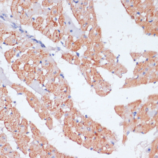
Immunohistochemistry analysis of paraffin-embedded Rat heart using β-arrestin1 Rabbit pAb at dilution of 1:100 (40x lens). Microwave antigen retrieval performed with 0.01M PBS Buffer (pH 7.2) prior to IHC staining.

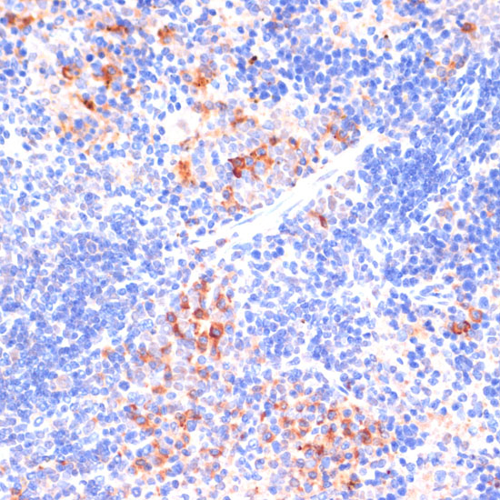
Immunohistochemistry analysis of paraffin-embedded Mouse spleen using β-arrestin1 Rabbit pAb at dilution of 1:100 (40x lens). Microwave antigen retrieval performed with 0.01M PBS Buffer (pH 7.2) prior to IHC staining.

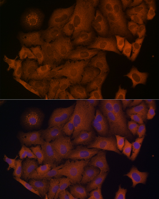
Immunofluorescence analysis of A-549 cells using β-arrestin1 Rabbit pAb at dilution of 1:100 (40x lens). Secondary antibody: Cy3-conjugated Goat anti-Rabbit IgG (H+L) at 1:500 dilution. Blue: DAPI for nuclear staining.

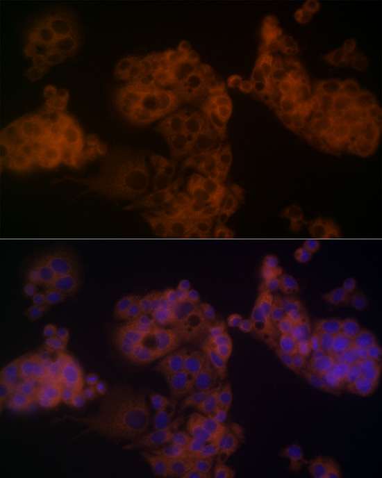
Immunofluorescence analysis of HepG2 cells using β-arrestin1 Rabbit pAb at dilution of 1:100 (40x lens). Secondary antibody: Cy3-conjugated Goat anti-Rabbit IgG (H+L) at 1:500 dilution. Blue: DAPI for nuclear staining.

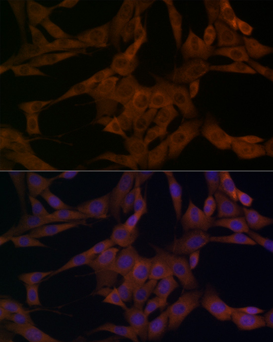
Immunofluorescence analysis of NIH/3T3 cells using β-arrestin1 Rabbit pAb at dilution of 1:100 (40x lens). Secondary antibody: Cy3-conjugated Goat anti-Rabbit IgG (H+L) at 1:500 dilution. Blue: DAPI for nuclear staining.

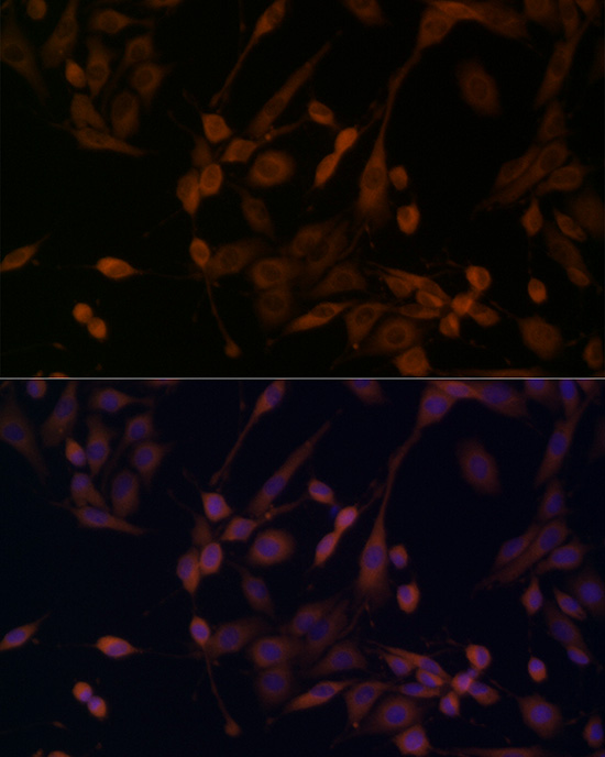
Immunofluorescence analysis of PC-12 cells using β-arrestin1 Rabbit pAb at dilution of 1:100 (40x lens). Secondary antibody: Cy3-conjugated Goat anti-Rabbit IgG (H+L) at 1:500 dilution. Blue: DAPI for nuclear staining.

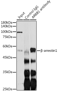
Immunoprecipitation analysis of 200 μg extracts of SH-SY5Y cells using 3 μg β-arrestin1 antibody, Western blot was performed from the immunoprecipitate using β-arrestin1 at a dilution of 1:1000.

No documents available.