Vimentin

Western blot analysis of extracts of various cell lines, using Vimentin antibody at 1:1000 dilution.
Secondary antibody: HRP Goat Anti-Rabbit IgG (H+L)  at 1:10000 dilution.
Lysates/proteins: 25μg per lane.
Blocking buffer: 3% nonfat dry milk in TBST.
Detection: ECL West Pico Plus.     
Exposure time: 1s.

Western blot analysis of extracts of various cell lines, using Vimentin antibody at 1:1000 dilution. Secondary antibody: HRP Goat Anti-Rabbit IgG (H+L) at 1:10000 dilution. Lysates/proteins: 25μg per lane. Blocking buffer: 3% nonfat dry milk in TBST. Detection: ECL West Pico Plus. Exposure time: 1s.

Catalog Number:AB-82376
Conjugate:Unconjugated
Size:100 ug
Concentration:1mg/ml
Host:Rabbit
Isotype:IgG
Clone:POLY
Immunogen:A synthetic peptide corresponding to a sequence within amino acids 367-466 of human Vimentin
Reactivity:Human, Mouse, Rat
Applications:Western Blot: 1:2000 - 1:20000 Immunohistochemistry (paraffin embedded tissues): HC-P 1:100 - 1:500 Immunofluorescence:1:50 - 1:200 Immunocytochemistry: 1:50 - 1:200 IP 1:500 - 1:1000
Molecular Weight:54kDa
Purification:Affinity purification
Form:Liquid
Synonyms:CTRCT30;HEL113;Vimentin;VIM;vimentin

Applications

Western Blot: 1:2000 - 1:20000 Immunohistochemistry (paraffin embedded tissues): HC-P 1:100 - 1:500 Immunofluorescence:1:50 - 1:200 Immunocytochemistry: 1:50 - 1:200 IP 1:500 - 1:1000

Immunogen

A synthetic peptide corresponding to a sequence within amino acids 367-466 of human Vimentin
Target Background

This gene encodes a type III intermediate filament protein. Intermediate filaments, along with microtubules and actin microfilaments, make up the cytoskeleton. The encoded protein is responsible for maintaining cell shape and integrity of the cytoplasm, and stabilizing cytoskeletal interactions. This protein is involved in neuritogenesis and cholesterol transport and functions as an organizer of a number of other critical proteins involved in cell attachment, migration, and signaling. Bacterial and viral pathogens have been shown to attach to this protein on the host cell surface. Mutations in this gene are associated with congenital cataracts in human patients.

Synonyms:CTRCT30;HEL113;Vimentin;VIM;vimentin

Storage

Store at -20℃. Avoid freeze / thaw cycles.

Buffer

PBS with 0.05% proclin300,0.05% BSA,50% glycerol,pH7.3.

Western blot analysis of extracts of various cell lines, using Vimentin antibody at 1:1000 dilution. Secondary antibody: HRP Goat Anti-Rabbit IgG (H+L) at 1:10000 dilution. Lysates/proteins: 25μg per lane. Blocking buffer: 3% nonfat dry milk in TBST. Detection: ECL West Pico Plus. Exposure time: 1s.

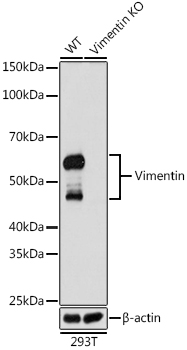
Western blot analysis of extracts from wild type (WT) and Vimentin knockout (KO) 293T cells, using Vimentin antibody at 1:1000 dilution. Secondary antibody: HRP Goat Anti-Rabbit IgG (H+L) at 1:10000 dilution. Lysates/proteins: 25μg per lane. Blocking buffer: 3% nonfat dry milk in TBST. Detection: ECL West Pico Plus. Exposure time: 1s.

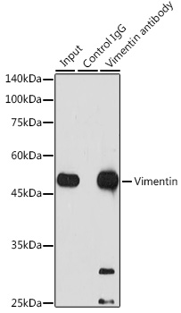
Immunoprecipitation analysis of 300 μg extracts of Jurkat cells using 3 μg Vimentin antibody . Western blot was performed from the immunoprecipitate using Vimentin antibody at a dilution of 1:1000.

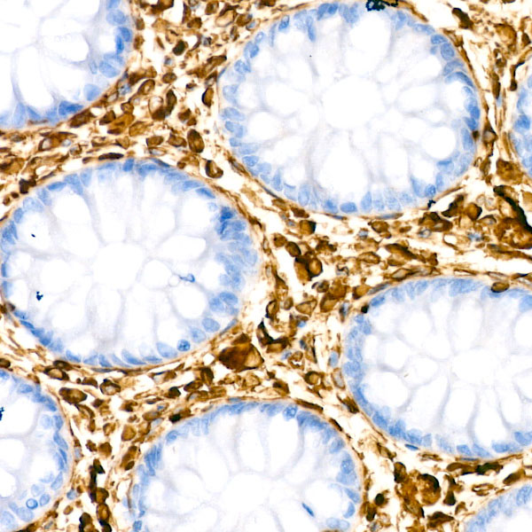
Immunohistochemistry analysis of paraffin-embedded human colon using [KO Validated] Vimentin antibody at dilution of 1:100 (40x lens).Perform microwave antigen retrieval with 10 mM PBS buffer pH 7.2 before commencing with IHC staining protocol.

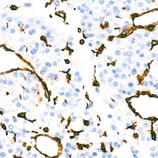
Immunohistochemistry analysis of paraffin-embedded human liver cancer using [KO Validated] Vimentin antibody at dilution of 1:100 (40x lens).Perform microwave antigen retrieval with 10 mM PBS buffer pH 7.2 before commencing with IHC staining protocol.

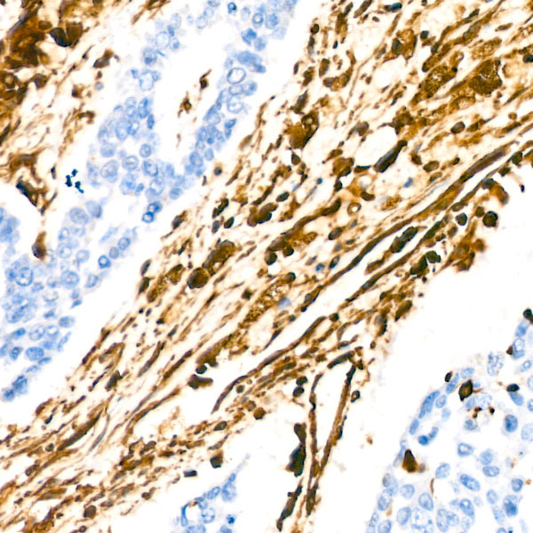
Immunohistochemistry analysis of paraffin-embedded human lung cancer using [KO Validated] Vimentin antibody at dilution of 1:100 (40x lens).Perform microwave antigen retrieval with 10 mM PBS buffer pH 7.2 before commencing with IHC staining protocol.

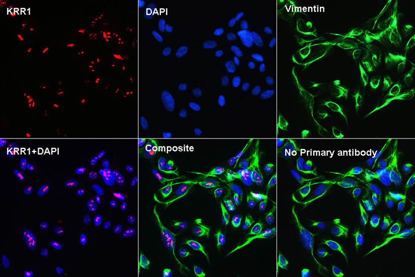
Confocal Immunofluorescence analysis of U2OS cells using KRR1 Rabbit pAb at dilution of 1:100 (40x lens)(red). Vimentin staining(green), DAPI for nuclear staining(blue).

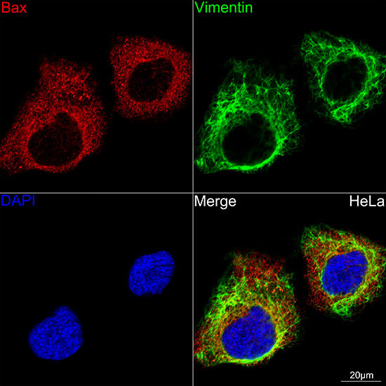
Confocal imaging of HeLa cells using [KO Validated] Vimentin antibody dilution 1:100) (Green). The cells were counterstained with Aurora B Rabbit mAb (dilution 1:800) (Red). DAPI was used for nuclear staining (blue). Objective: 60x.

No documents available.