VMAT1

Figure 1. Western blot analysis of SLC18A1 using anti-
SLC18A1 antibody.      
Electrophoresis was performed on a 5-20% SDS-PAGE gel
at 70V (Stacking gel) / 90V (Resolving gel) for 2-3 hours. The
sample well of each lane was loaded with 50ug of sample
under reducing conditions.
Lane 1: human Hela whole cell lysates,
Lane 2: human A549 whole cell lysates,
Lane 3: human U-87MG whole cell lysates,
Lane 4: human A431 whole cell lysates,
Lane 5: human HL-60 cell lysates,
Lane 6: human K562 cell lysates.
After Electrophoresis, proteins were transferred to a
Nitrocellulose membrane at 150mA for 50-90 minutes.
Blocked the membrane with 5% Non-fat Milk/ TBS for 1.5
hour at RT. The membrane was incubated with rabbit anti-
SLC18A1 antigen affinity purified polyclonal antibody
at 0.5 mug/mL overnight at 4°C, then washed with TBS-0.1%Tween 3 times with 5 minutes each
and probed with a goat anti-rabbit IgG-HRP secondary
antibody at a dilution of 1:5000 for 1.5 hour at RT. The
signal is developed using an Enhanced Chemiluminescent
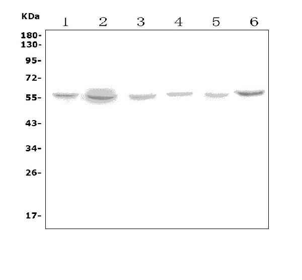
detection (ECL) kit with Tanon 5200  system. A specific band was detected for SLC18A1 at approximately 56KD. The expected band size for SLC18A1 is  at 56KD.

Figure 1. Western blot analysis of SLC18A1 using anti- SLC18A1 antibody. Electrophoresis was performed on a 5-20% SDS-PAGE gel at 70V (Stacking gel) / 90V (Resolving gel) for 2-3 hours. The sample well of each lane was loaded with 50ug of sample under reducing conditions. Lane 1: human Hela whole cell lysates, Lane 2: human A549 whole cell lysates, Lane 3: human U-87MG whole cell lysates, Lane 4: human A431 whole cell lysates, Lane 5: human HL-60 cell lysates, Lane 6: human K562 cell lysates. After Electrophoresis, proteins were transferred to a Nitrocellulose membrane at 150mA for 50-90 minutes. Blocked the membrane with 5% Non-fat Milk/ TBS for 1.5 hour at RT. The membrane was incubated with rabbit anti- SLC18A1 antigen affinity purified polyclonal antibody at 0.5 mug/mL overnight at 4°C, then washed with TBS-0.1%Tween 3 times with 5 minutes each and probed with a goat anti-rabbit IgG-HRP secondary antibody at a dilution of 1:5000 for 1.5 hour at RT. The signal is developed using an Enhanced Chemiluminescent detection (ECL) kit with Tanon 5200 system. A specific band was detected for SLC18A1 at approximately 56KD. The expected band size for SLC18A1 is at 56KD.

Catalog Number:AB-82380
Conjugate:Unconjugated
Size:100 ug
Concentration:1mg/ml
Host:Rb
Isotype:IgG
Clone:POLY
Immunogen:E.coli-derived human VMAT1/SLC18A1 recombinant protein (Position: M1-E510).
Reactivity:Hu
Applications:Western blot: 0.25-0.5mug/ml Immunohistochemistry(Paraffin-embedded Section): 0.5-1mug/ml Flow Cytometry: 1-3mug/1x106 cells Direct ELISA: 0.1-0.5mug/ml
Purification:Aff. Pur.
Form:Liquid

Applications

Western blot: 0.25-0.5mug/ml Immunohistochemistry(Paraffin-embedded Section): 0.5-1mug/ml Flow Cytometry: 1-3mug/1x106 cells Direct ELISA: 0.1-0.5mug/ml

Immunogen

E.coli-derived human VMAT1/SLC18A1 recombinant protein (Position: M1-E510).
Target Background

Chromaffin granule amine transporter is a protein that in humans is encoded by the SLC18A1 gene. This gene is mapped to 8p21.3. The vesicular monoamine transporter acts to accumulate cytosolic monoamines into vesicles, using the proton gradient maintained across the vesicular membrane. Its proper function is essential to the correct activity of the monoaminergic systems that have been implicated in several human neuropsychiatric disorders. The transporter is a site of action of important drugs, including reserpine and tetrabenazine.

Storage

At -20°C for one year. It can also be aliquotted and stored frozen at -20°C for a longer time. Avoid repeated freezing and

Buffer

Each vial contains 4mg Trehalose, 0.9mg NaCl, 0.2mg Na2HPO4, 0.05mg NaN3.

Figure 1. Western blot analysis of SLC18A1 using anti- SLC18A1 antibody. Electrophoresis was performed on a 5-20% SDS-PAGE gel at 70V (Stacking gel) / 90V (Resolving gel) for 2-3 hours. The sample well of each lane was loaded with 50ug of sample under reducing conditions. Lane 1: human Hela whole cell lysates, Lane 2: human A549 whole cell lysates, Lane 3: human U-87MG whole cell lysates, Lane 4: human A431 whole cell lysates, Lane 5: human HL-60 cell lysates, Lane 6: human K562 cell lysates. After Electrophoresis, proteins were transferred to a Nitrocellulose membrane at 150mA for 50-90 minutes. Blocked the membrane with 5% Non-fat Milk/ TBS for 1.5 hour at RT. The membrane was incubated with rabbit anti- SLC18A1 antigen affinity purified polyclonal antibody at 0.5 mug/mL overnight at 4°C, then washed with TBS-0.1%Tween 3 times with 5 minutes each and probed with a goat anti-rabbit IgG-HRP secondary antibody at a dilution of 1:5000 for 1.5 hour at RT. The signal is developed using an Enhanced Chemiluminescent detection (ECL) kit with Tanon 5200 system. A specific band was detected for SLC18A1 at approximately 56KD. The expected band size for SLC18A1 is at 56KD.

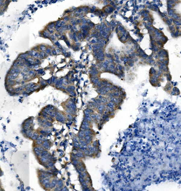
Figure 2. IHC analysis of SLC18A1 using anti-SLC18A1 antibody. SLC18A1 was detected in paraffin-embedded section of human pancreatic cancer tissue. Heat mediated antigen retrieval was performed in EDTA buffer (pH8.0, epitope retrieval solution). The tissue section was blocked with 10% goat serum. The tissue section was then incubated with 1μg/ml rabbit anti-SLC18A1 Antibody overnight at 4°C. Biotinylated goat anti-rabbit IgG was used as secondary antibody and incubated for 30 minutes at 37°C. The tissue section was developed using Strepavidin-Biotin-Complex (SABC) with DAB as the chromogen.

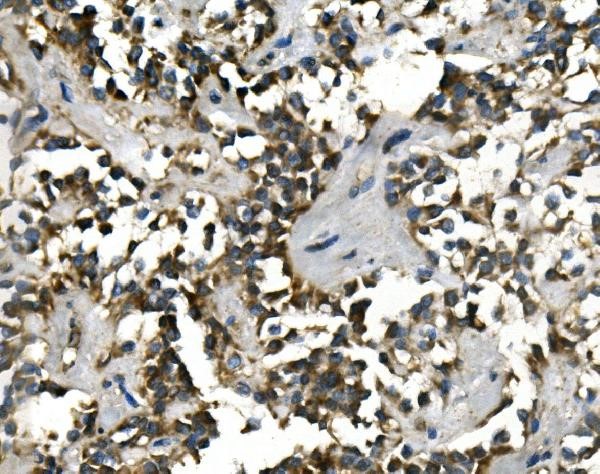
Figure 3. IHC analysis of SLC18A1 using anti-SLC18A1 Antibody. SLC18A1 was detected in paraffin-embedded section of human rectal cancer tissue. Heat mediated antigen retrieval was performed in EDTA buffer (pH8.0, epitope retrieval solution). The tissue section was blocked with 10% goat serum. The tissue section was then incubated with 1mug/ml rabbit anti-SLC18A1 Antibody overnight at 4°C. Biotinylated goat anti-rabbit IgG was used as secondary antibody and incubated for 30 minutes at 37°C. The tissue section was developed using Strepavidin-Biotin-Complex(SABC) with DAB as the chromogen.

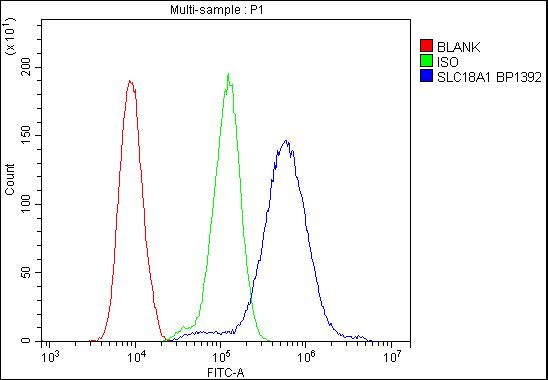
Figure 4. Flow Cytometry analysis of HL-60 cells using anti-SLC18A1 antibody Overlay histogram showing HL-60 cells stained with (Blue line).The cells were blocked with 10% normal goat serum. And then incubated with rabbit anti- SLC18A1 Antibody 1mug/1x106 cells) for 30 min at 20°C. DyLight®488 conjugated goat anti-rabbit IgG was used as secondary antibody for 30 minutes at 20°C. Isotype control antibody (Green line) was rabbit IgG (1mug/1x106) used under the same conditions. Unlabelled sample (Red line) was also used as a control.

No documents available.