
VPS26A

Western blot analysis of extracts of various cell lines, 
using VPS26A antibody at 1:1000
dilution.
Secondary antibody: HRP Goat Anti-Rabbit IgG
(H+L) at 1:10000 dilution.
Lysates/proteins: 25ug per lane.
Blocking buffer: 3% nonfat dry milk in TBST.
Detection: ECL West Pico Plus.   
Exposure time: 5s.

Western blot analysis of extracts of various cell lines, using VPS26A antibody at 1:1000 dilution. Secondary antibody: HRP Goat Anti-Rabbit IgG (H+L) at 1:10000 dilution. Lysates/proteins: 25ug per lane. Blocking buffer: 3% nonfat dry milk in TBST. Detection: ECL West Pico Plus. Exposure time: 5s.

Catalog Number:AB-84398
Conjugate:Unconj.
Size:100ug
Concentration:1mg/ml
Host:Rb
Isotype:100 ug
Clone:POLY
Immunogen:Recombinant fusion protein containing a sequence corresponding to amino acids 208-327 of human VPS26A.
Reactivity:Hu, Ms, Rt
Applications:Western Blot: 1:500 - 1:2000 Immunofluorescence: 1:50 - 1:200
Molecular Weight:38kDa
Purification:Aff. Pur.
Form:Liquid
Synonyms:VPS26A;HB58;Hbeta58;PEP8A;VPS26

Applications

Western Blot: 1:500 - 1:2000 Immunofluorescence: 1:50 - 1:200

Immunogen

Recombinant fusion protein containing a sequence corresponding to amino acids 208-327 of human VPS26A.

Target Background

This gene belongs to a group of vacuolar protein sorting (VPS) genes. The encoded protein is a component of a large multimeric complex, termed the retromer complex, involved in retrograde transport of proteins from endosomes to the trans-Golgi network. The close structural similarity between the yeast and human proteins that make up this complex suggests a similarity in function. Expression studies in yeast and mammalian cells indicate that this protein interacts directly with VPS35, which serves as the core of the retromer complex. Alternative splicing results in multiple transcript variants encoding different isoforms.

Synonyms:VPS26A;HB58;Hbeta58;PEP8A;VPS26

Storage

Store at -20℃. Avoid freeze / thaw cycles.

Buffer

PBS with 0.02% sodium azide,50% glycerol,pH7.3.

Western blot analysis of extracts of various cell lines, using VPS26A antibody at 1:1000 dilution. Secondary antibody: HRP Goat Anti-Rabbit IgG (H+L) at 1:10000 dilution. Lysates/proteins: 25ug per lane. Blocking buffer: 3% nonfat dry milk in TBST. Detection: ECL West Pico Plus. Exposure time: 5s.

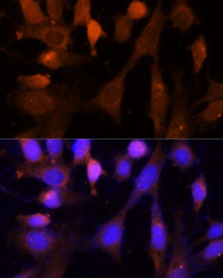
Immunofluorescence analysis of NIH-3T3 cells using VPS26A Polyclonal Antibody at dilution of 1:100 (40x lens). Blue: DAPI for nuclear staining.

No documents available.