WNT10B

Western blot analysis of extracts of
HeLa cells, using WNT10B antibody
at 1:1000 dilution.
Secondary antibody: HRP Goat Anti-
Rabbit IgG (H+L) at 1:10000
dilution.
Lysates/proteins: 25ug per lane.
Blocking buffer: 3% nonfat dry milk in
TBST.
Detection: ECL West Pico Plus. 
Exposure time: 10s.

Western blot analysis of extracts of HeLa cells, using WNT10B antibody at 1:1000 dilution. Secondary antibody: HRP Goat Anti- Rabbit IgG (H+L) at 1:10000 dilution. Lysates/proteins: 25ug per lane. Blocking buffer: 3% nonfat dry milk in TBST. Detection: ECL West Pico Plus. Exposure time: 10s.

Catalog Number:AB-84168
Size:100 ug
Concentration:1mg/ml
Host:Rb
Isotype:IgG
Clone:POLY
Immunogen:Recombinant Protein of human WNT10B
Reactivity:Hu, Ms, Rt
Applications:Western Blot: 1:250 - 1:1000 Immunohistochemistry (paraffin-embedded tissues):1:25 - 1:100 Immunofluorescence: 1:25 - 1:100
Molecular Weight:43kDa
Purification:Aff. Pur.
Form:Liquid
Synonyms:WNT10B; SHFM6; STHAG8; WNT-12; protein Wnt-10b

Applications

Western Blot: 1:250 - 1:1000 Immunohistochemistry (paraffin-embedded tissues):1:25 - 1:100 Immunofluorescence: 1:25 - 1:100

Immunogen

Recombinant Protein of human WNT10B
Target Background

The WNT gene family consists of structurally related genes which encode secreted signaling proteins. These proteins have been implicated in oncogenesis and in several developmental processes, including regulation of cell fate and patterning during embryogenesis. This gene is a member of the WNT gene family. It may be involved in breast cancer, and its protein signaling is likely a molecular switch that governs adipogenesis. This protein is 96% identical to the mouse Wnt10b protein at the amino acid level. This gene is clustered with another family member, WNT1, in the chromosome 12q13 region.

Synonyms:WNT10B; SHFM6; STHAG8; WNT-12; protein Wnt-10b

Storage

Store at -20℃. Avoid freeze / thaw cycles.

Buffer

PBS with 0.02% sodium azide, pH7.3.

Western blot analysis of extracts of HeLa cells, using WNT10B antibody at 1:1000 dilution. Secondary antibody: HRP Goat Anti- Rabbit IgG (H+L) at 1:10000 dilution. Lysates/proteins: 25ug per lane. Blocking buffer: 3% nonfat dry milk in TBST. Detection: ECL West Pico Plus. Exposure time: 10s.

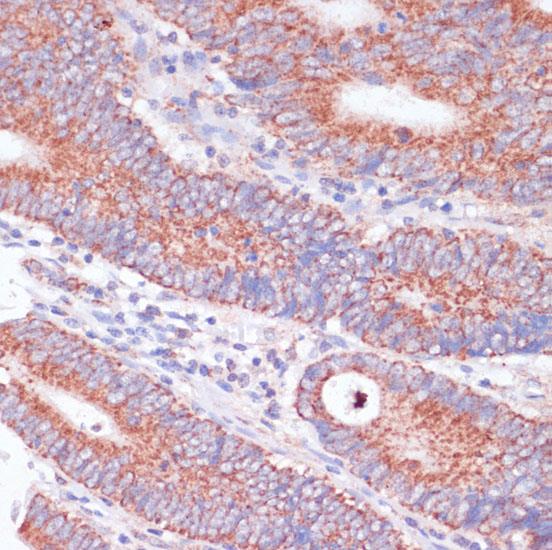
Immunohistochemistry of paraffin-embedded human colon carcinoma using WNT10B antibody at dilution of 1:100 (40x lens).

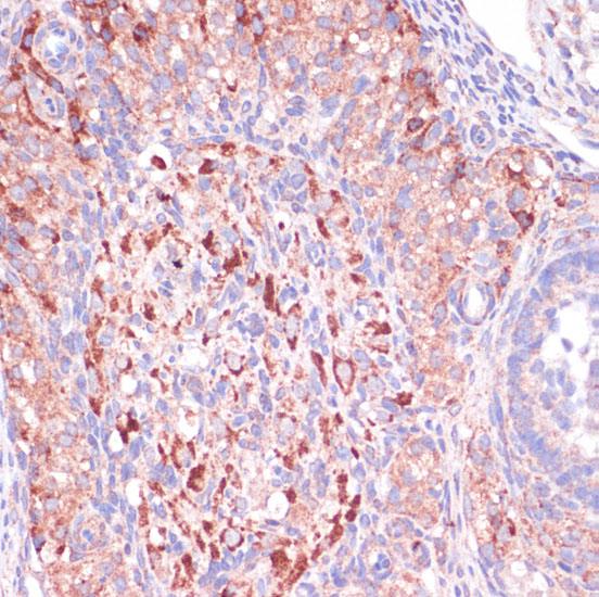
Immunohistochemistry of paraffin-embedded rat ovary using WNT10B antibody at dilution of 1:100 (40x lens).

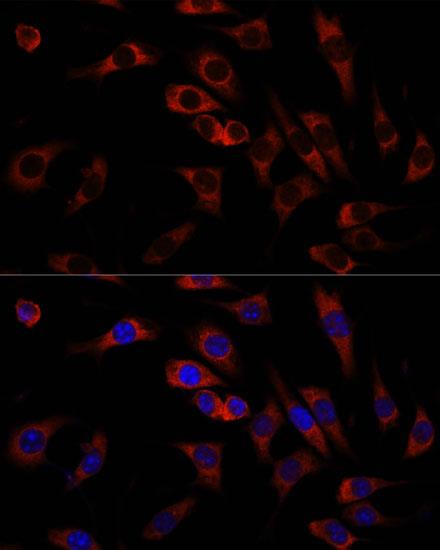
Immunofluorescence analysis of C6 cells using WNT10B antibody at dilution of 1:100. Blue: DAPI for nuclear staining.

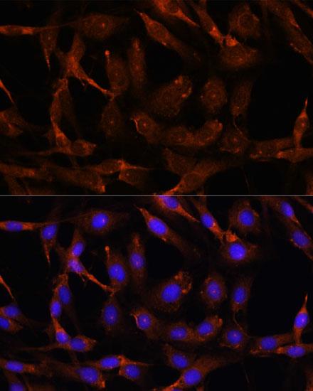
Immunofluorescence analysis of L929 cells using WNT10B antibody at dilution of 1:100. Blue: DAPI for nuclear staining.

No documents available.