
XIAP

Western blot analysis of extracts of various
 cell lines, using XIAP antibody at 1:1000 dilution.
Secondary antibody: HRP Goat Anti-Rabbit IgG (H+L) at 1:10000 dilution. Lysates/proteins: 25ug per lane.
Blocking buffer: 3% nonfat dry milk in TBST.
Detection: ECL West Pico Plus.   
Exposure time: 10s.

Western blot analysis of extracts of various cell lines, using XIAP antibody at 1:1000 dilution. Secondary antibody: HRP Goat Anti-Rabbit IgG (H+L) at 1:10000 dilution. Lysates/proteins: 25ug per lane. Blocking buffer: 3% nonfat dry milk in TBST. Detection: ECL West Pico Plus. Exposure time: 10s.

Catalog Number:AB-84345
Size:100μg
Concentration:1mg/ml
Host:Rb
Isotype:IgG
Clone:POLY
Immunogen:Recombinant fusion protein containing a sequence corresponding to amino acids 1-200 of human XIAP.
Reactivity:Hu,Ms,Rt
Applications:Western Blot: 1:500 - 1:2000 Immunofluorescence: 1:50 - 1:200
Molecular Weight:51kDa
Purification:Aff. Pur.
Form:Liquid
Synonyms:XIAP;API3;BIRC4;IAP-3;ILP1;MIHA;XLP2;hIAP-3;hIAP3

Applications

Western Blot: 1:500 - 1:2000 Immunofluorescence: 1:50 - 1:200

Immunogen

Recombinant fusion protein containing a sequence corresponding to amino acids 1-200 of human XIAP.
Target Background

This gene encodes a protein that belongs to a family of apoptotic suppressor proteins. Members of this family share a conserved motif termed, baculovirus IAP repeat, which is necessary for their anti-apoptotic function. This protein functions through binding to tumor necrosis factor receptor-associated factors TRAF1 and TRAF2 and inhibits apoptosis induced by menadione, a potent inducer of free radicals, and interleukin 1- beta converting enzyme. This protein also inhibits at least two members of the caspase family of cell-death proteases, caspase-3 and caspase-7. Mutations in this gene are the cause of X-linked lymphoproliferative syndrome. Alternate splicing results in multiple transcript variants. Pseudogenes of this gene are found on chromosomes 2 and 11.

Synonyms:XIAP;API3;BIRC4;IAP-3;ILP1;MIHA;XLP2;hIAP-3;hIAP3

Storage

Store at -20℃. Avoid freeze / thaw cycles.

Buffer

PBS with 0.02% sodium azide,50% glycerol,pH7.3.

Western blot analysis of extracts of various cell lines, using XIAP antibody at 1:1000 dilution. Secondary antibody: HRP Goat Anti-Rabbit IgG (H+L) at 1:10000 dilution. Lysates/proteins: 25ug per lane. Blocking buffer: 3% nonfat dry milk in TBST. Detection: ECL West Pico Plus. Exposure time: 10s.

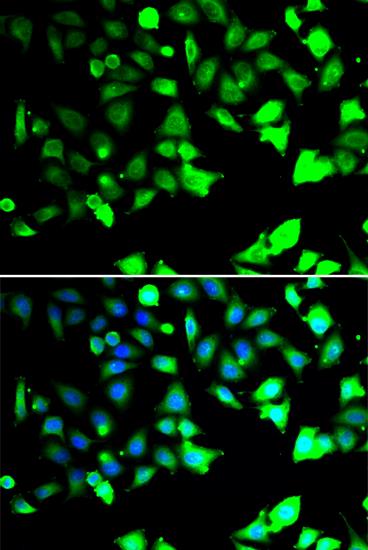
Immunofluorescence analysis of HeLa cells using XIAP antibody. Blue: DAPI for nuclear staining.

No documents available.