YAP

Western blot analysis of extracts of
various cell lines, using YAP1 antibody
at 1:1000 dilution. Secondary antibody:
HRP Goat Anti- Rabbit IgG (H+L)at
1:10000 dilution.
Lysates/proteins: 25ug per lane.
Blocking buffer: 3% nonfat dry milk in
TBST.
Detection: ECL West Pico Plus
Exposure time: 10s.

Western blot analysis of extracts of various cell lines, using YAP1 antibody at 1:1000 dilution. Secondary antibody: HRP Goat Anti- Rabbit IgG (H+L)at 1:10000 dilution. Lysates/proteins: 25ug per lane. Blocking buffer: 3% nonfat dry milk in TBST. Detection: ECL West Pico Plus Exposure time: 10s.

Catalog Number:MAB-94469
Size:100μg
Concentration:1mg/ml
Host:Rb
Isotype:IgG
Clone:D8H1X
Reactivity:Hu,Ms,Rt
Applications:Western blotting 1:1000 Immunoprecipitation 1:20-1:50 Immunohistochemistry (Paraffin) Unmasking buffer: Immunofluorescence IF Size: 100 ug Concentration: 1mg/ml Clone: D8H1X 1:50-1:200 1:50-1:200
Molecular Weight:65-75 kDa
Purification:Monoclonal antibody is produced by immunizing animals with recombinant protein specific to the carboxy terminus of human YAP protein. The epitope corresponds to a region surrounding Pro435 of human YAP isoform 1. This sequence region is 100% conserved among all known isoforms of human YAP protein.
Form:Liquid

Applications

Western blotting 1:1000 Immunoprecipitation 1:20-1:50 Immunohistochemistry (Paraffin) Unmasking buffer: Immunofluorescence IF Size: 100 ug Concentration: 1mg/ml Clone: D8H1X 1:50-1:200 1:50-1:200

Target Background

YAP (Yes-associated protein, YAP65) was identified based on its ability to associate with the SH3 domain of Yes. It also binds to other SH3 domain-containing proteins such as Nck, Crk, Src, and Abl (1). In addition to the SH3 binding motif, YAP contains a PDZ interaction motif, a coiled-coil domain, and WW domains (2-4). While initial studies of YAP all pointed towards a role in anchoring and targeting to specific subcellular compartments, subsequent studies showed that YAP is a transcriptional coactivator by virtue of its WW domain interacting with the PY motif (PPxY) of the transcription factor PEBP2 and other transcription factors (5,6). In its capacity as a transcriptional coactivator, YAP is now widely recognized as a central mediator of the Hippo Pathway, which plays a fundamental and widely conserved role in regulating tissue growth and organ size. Upon phosphorylation at Ser127 by LATS1/2 kinases, YAP translocates to the cytoplasm, where it is sequestered through association with 14-3-3 proteins in an Akt-dependent manner (6-8).YAP (D8H1X) XP® Rabbit mAb recognizes endogenous levels of total YAP protein.

Buffer

PBS with 0.02%sodium azide, 50% glycerol, pH7.3.

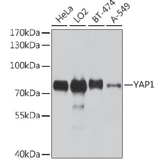
Western blot analysis of extracts of various cell lines, using YAP1 antibody at 1:1000 dilution. Secondary antibody: HRP Goat Anti- Rabbit IgG (H+L)at 1:10000 dilution. Lysates/proteins: 25ug per lane. Blocking buffer: 3% nonfat dry milk in TBST. Detection: ECL West Pico Plus Exposure time: 10s.

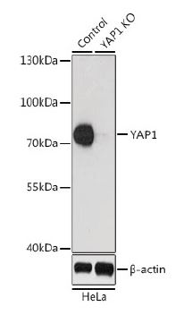
Western blot analysis of extracts from normal (control) and YAP1 knockout (KO) HeLa cells, using YAP1 antibody at 1:1000 dilution. Secondary antibody: HRP Goat Anti- Rabbit IgG (H+L) at 1:10000 dilution. Lysates/proteins: 25ug per lane. Blocking buffer: 3% nonfat dry milk in TBST. Detection: ECL Enhanced Kit. Exposure time: 90s.

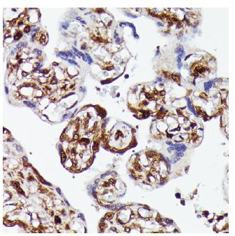
Immunohistochemistry of paraffinembedded human placenta using YAP1 antibody at dilution of 1:100 (40x lens).

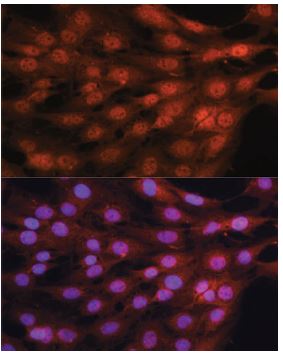
Immunofluorescence analysis of C6 cells using YAP1 antibody at dilution of 1:100. Blue: DAPI for nuclear staining.

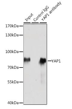
Immunoprecipitation analysis of 200ug extracts of HeLa cells, using 3 ug YAP1 antibody. Western blot was performed from the immunoprecipitate using YAP1 antibody (at a dilition of 1:1000.

No documents available.