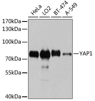
Western blot analysis of extracts of various cell lines, using YAP1 antibody  at 1:1000 dilution.
Secondary antibody: HRP Goat Anti-Rabbit IgG (H+L) at 1:10000 dilution.
Lysates/proteins: 25ug per lane.
Blocking buffer: 3% nonfat dry milk in TBST.
Detection: ECL West Pico Plus. 
Exposure time: 10s.

Western blot analysis of extracts of various cell lines, using YAP1 antibody at 1:1000 dilution. Secondary antibody: HRP Goat Anti-Rabbit IgG (H+L) at 1:10000 dilution. Lysates/proteins: 25ug per lane. Blocking buffer: 3% nonfat dry milk in TBST. Detection: ECL West Pico Plus. Exposure time: 10s.

Catalog Number:AB-84335
Size:100μg
Concentration:1mg/ml
Host:Rb
Isotype:IgG
Clone:POLY
Immunogen:Recombinant fusion protein containing a sequence corresponding to amino acids 155-504 of human YAP1.
Reactivity:Hu,Ms,Rt
Applications:Western Blot: 1:500 - 1:2000 Immunohistochemistry: 1:50 - 1:200 Immunofluorescence: 1:50 - 1:200 Immunoprecipitation: 1:20 - 1:50
Molecular Weight:72kDa
Purification:Aff. Pur.
Form:Liquid
Synonyms:COB1;YAP;YAP2;YAP65;YKI;YAP1

Applications

Western Blot: 1:500 - 1:2000 Immunohistochemistry: 1:50 - 1:200 Immunofluorescence: 1:50 - 1:200 Immunoprecipitation: 1:20 - 1:50

Immunogen

Recombinant fusion protein containing a sequence corresponding to amino acids 155-504 of human YAP1.

Target Background

This gene encodes a downstream nuclear effector of the Hippo signaling pathway which is involved in development, growth, repair, and homeostasis. This gene is known to play a role in the development and progression of multiple cancers as a transcriptional regulator of this signaling pathway and may function as a potential target for cancer treatment. Alternative splicing results in multiple transcript variants encoding different isoforms.

Synonyms:COB1;YAP;YAP2;YAP65;YKI;YAP1

Storage

Store at -20°C. Avoid freeze/ thaw cycles.

Buffer

PBS with 0.02%sodium azide, 50% glycerol, pH7.3.

Western blot analysis of extracts of various cell lines, using YAP1 antibody at 1:1000 dilution. Secondary antibody: HRP Goat Anti-Rabbit IgG (H+L) at 1:10000 dilution. Lysates/proteins: 25ug per lane. Blocking buffer: 3% nonfat dry milk in TBST. Detection: ECL West Pico Plus. Exposure time: 10s.

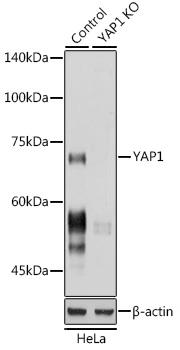
Western blot analysis of extracts from normal (control) and [KO Validated] YAP1 Rabbit pAb knockout (KO) HeLa cells, using [KO Validated] YAP1 Rabbit pAb antibody at 1:1000 dilution. Secondary antibody: HRP Goat Anti-Rabbit IgG (H+L) at 1:10000 dilution. Lysates/proteins: 25ug per lane. Blocking buffer: 3% nonfat dry milk in TBST. Detection: ECL West Pico Plus. Exposure time: 1s.

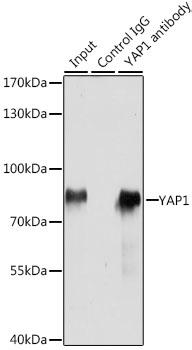
Immunoprecipitation analysis of 200ug extracts of HeLa cells, using 3 ug YAP1 antibody . Western blot was performed from the immune precipitate using YAP1 antibody at a dilition of 1:1000.

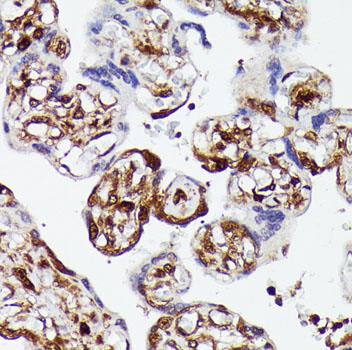
Immunohistochemistry of paraffin embedded human placenta using YAP1 antibody at dilution of 1:100 (40x lens).

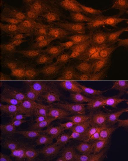
Immunofluorescence analysis of C6 cells using YAP1 antibody at dilution of 1:100. Blue: DAPI for nuclear staining.

No documents available.